AI Breakthrough: Simulating Brain Activity from Connectome Data
3 Sources
3 Sources
[1]
New AI method simulates neuronal activity from connectome data
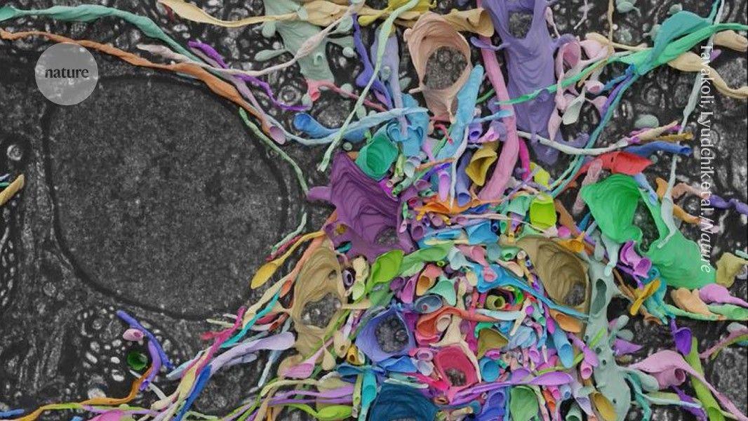
Howard Hughes Medical InstituteSep 12 2024 With maps of the connections between neurons and artificial intelligence methods, researchers can now do what they never thought possible: predict the activity of individual neurons without making a single measurement in a living brain. For decades, neuroscientists have spent countless hours in the lab painstakingly measuring the activity of neurons in living animals to tease out how the brain enables behavior. These experiments have yielded groundbreaking insights into how the brain works, but they have only scratched the surface, leaving much of the brain unexplored. Now, researchers are using artificial intelligence and the connectome - a map of neurons and their connections created from brain tissue - to predict the role of neurons in the living brain. Using only information about the connectivity of a neural circuit gleaned from the fruit fly visual system connectome and a guess at what the circuit is supposed to do, researchers created an AI simulation of the fruit fly visual system that can predict the activity of every neuron in the circuit. We now have a computational method for turning measurements of the connectome into predictions of neural activity and brain function, without first starting with difficult-to-acquire measurements of neural activity for every neuron." Srini Turaga, Janelia Group Leader, senior author on the new research The team of scientists from HHMI's Janelia Research Campus and the University of Tübingen used the connectome to build a detailed deep mechanistic network simulation of the fly visual system, where each neuron and synapse in the model corresponds to a real neuron and synapse in the brain. Although they didn't know the dynamics of every neuron and synapse, data from the connectome allowed the team to use deep learning methods to infer these unknown parameters. They combined this information with knowledge about the circuit's goal: motion detection. "At that point, everything fell into place, and we could finally figure out if this connectome-constrained model gives us a good model of the brain," says Janne Lappalainen, a PhD student at the University of Tübingen who led the research. The new model predicts the neural activity produced by 64 neuron types in the fruit fly visual system in response to visual input and accurately reproduces more than two dozen experimental studies performed over the past two decades. By enabling researchers to predict the activity of individual neurons using only the connectome, the new work has the potential to transform how neuroscientists generate and test hypotheses about how the brain works. In principle, scientists can now use the model to simulate any experiment and generate detailed predictions that can be tested in the lab. The new research provides more than 450 pages of predictions gleaned from the new model, including identification of cells not known to be involved in motion detection previously, which can now be examined in living flies. The group's work provides a strategy for turning the wealth of connectome data being generated by Janelia and other research institutions into advanced understanding of the living brain, according to the researchers. "There is a big gap between the static snapshot of the connectome and the dynamics of real-life computation in the living brain, and the question was, can we bridge that gap in a model? This paper, for the specific example of the fruit fly, shows a strategy for bridging that gap," says Jakob Macke, a senior author on the paper and a professor at the University of Tübingen. The new research showcases the potential of AI to accelerate scientific discovery. HHMI recently announced AI@HHMI, a $500 million investment over the next 10 years to support artificial intelligence-driven projects in the life sciences. Howard Hughes Medical Institute Journal reference: Lappalainen, J. K., et al. (2024). Connectome-constrained networks predict neural activity across the fly visual system. Nature. doi.org/10.1038/s41586-024-07939-3.
[2]
Combining the power of AI and the connectome to predict brain cell activity
With maps of the connections between neurons and artificial intelligence methods, researchers can now do what they never thought possible: predict the activity of individual neurons without making a single measurement in a living brain. For decades, neuroscientists have spent countless hours in the lab painstakingly measuring the activity of neurons in living animals to tease out how the brain enables behavior. These experiments have yielded groundbreaking insights into how the brain works, but they have only scratched the surface, leaving much of the brain unexplored. Now, researchers are using artificial intelligence and the connectome -- a map of neurons and their connections created from brain tissue -- to predict the role of neurons in the living brain. Their paper has been published in the journal Nature. Using only information about the connectivity of a neural circuit gleaned from the fruit fly visual system connectome and a guess at what the circuit is supposed to do, researchers created an AI simulation of the fruit fly visual system that can predict the activity of every neuron in the circuit. "We now have a computational method for turning measurements of the connectome into predictions of neural activity and brain function, without first starting with difficult-to-acquire measurements of neural activity for every neuron," says Janelia Group Leader Srini Turaga, a senior author on the new research. The team of scientists from HHMI's Janelia Research Campus and the University of Tübingen used the connectome to build a detailed deep mechanistic network simulation of the fly visual system, where each neuron and synapse in the model corresponds to a real neuron and synapse in the brain. Although they didn't know the dynamics of every neuron and synapse, data from the connectome allowed the team to use deep learning methods to infer these unknown parameters. They combined this information with knowledge about the circuit's goal: motion detection. "At that point, everything fell into place, and we could finally figure out if this connectome-constrained model gives us a good model of the brain," says Janne Lappalainen, a Ph.D. student at the University of Tübingen who led the research. The new model predicts the neural activity produced by 64 neuron types in the fruit fly visual system in response to visual input and accurately reproduces more than two dozen experimental studies performed over the past two decades. By enabling researchers to predict the activity of individual neurons using only the connectome, the new work has the potential to transform how neuroscientists generate and test hypotheses about how the brain works. In principle, scientists can now use the model to simulate any experiment and generate detailed predictions that can be tested in the lab. The new research provides more than 450 pages of predictions gleaned from the new model, including identification of cells not known to be involved in motion detection previously, which can now be examined in living flies. The group's work provides a strategy for turning the wealth of connectome data being generated by Janelia and other research institutions into advanced understanding of the living brain, according to the researchers. "There is a big gap between the static snapshot of the connectome and the dynamics of real-life computation in the living brain, and the question was, can we bridge that gap in a model? This paper, for the specific example of the fruit fly, shows a strategy for bridging that gap," says Jakob Macke, a senior author on the paper and a professor at the University of Tübingen.
[3]
Fly-brain connectome helps to make predictions about neural activity
Neural activity in the brain depends on how neuronal cells are connected, as well as the dynamics of individual neurons and synaptic connections in the circuit. Highly detailed connectomes (wiring diagrams) can now be constructed post-mortem using 3D electron microscopy. However, making equally detailed measurements of the dynamic properties of each neuron and synapse in a circuit is not yet possible. As a result, researchers have not been able to simulate a neural network and predict neural activity to understand brain function. This shortcoming has led many to question the usefulness of connectomic measurements, especially given the considerable effort involved in making them. How can we infer the dynamics of neurons and synapses to accurately predict neural activity from a wiring diagram? Our key insight was that we could deduce neural-activity dynamics by using knowledge of the computation performed by the brain. Such computation results from collective patterns of the activity of networks of neurons, in response to inputs. For example, motion-sensitive neurons in the fly visual system increase their firing in response to moving stimuli, and this enables flies to be highly sensitive to seeing motion. We constructed a neural-network simulation of the fly visual system with the same wiring diagram as that of the real fly brain, and incorporated free parameters that correspond to the unknown dynamic properties of each neuron and synapse in the network. We then used an artificial intelligence (AI) method called deep learning to search for parameters that enabled neurons in the simulated network to detect visual motion. We compared the neural activity predicted by our simulation with experimental recordings of individual neurons, taken from 26 previous studies encompassing more than 20 years (Fig. 1a). Our simulations predicted the neural activity of living fly brains with high accuracy (Fig. 1b). We could predict not only which neurons of the visual system underpinned motion detection, but also the mechanisms of this visual computation -- and others -- at single-neuron resolution. Our approach provides a way to generate detailed hypotheses about how the brain works using wiring diagrams, and allows us to make many experimentally testable predictions. Understanding the function of a brain region, cell type or neuron has conventionally required researchers to make challenging experimental measurements in a living brain. This is because many theories of brain function do not clearly incorporate specific components of the brain, and cannot therefore make detailed predictions. By contrast, our models have a one-to-one correspondence between simulated and real neurons. This enables us to make intricate predictions about the function of brain regions, cell types and neurons, even without measurements of neural activity from a living brain. The present work represents a comprehensive and detailed model of a biological visual system. Popular AI architectures called convolutional neural networks were inspired by a relatively crude understanding of biological vision. It is intriguing to ask what new principles we can learn from the compact and efficient fly visual system, and whether this model could inform progress in machine vision. Our study shows the impressive degree to which a simulation that models the wiring diagram in detail, but simplifies the dynamics of individual neurons and synapses, can predict brain function. Future models could incorporate features of biological networks -- such as synaptic plasticity, neuromodulation, electrical synapses and glial cells -- to enable researchers to understand brain function in greater detail and over longer timescales. Connectomes of the entire fly brain and nerve cord are now available. In parallel, our group has developed a biomechanical simulation of the fly body that can model realistic behaviour. Together, these measurements and models will enable embodied simulations of the entire fly nervous system, leading to a mechanistic simulation of the whole brain and body in exceptional detail. -- Srinivas C. Turaga is at the Janelia Research Campus, Howard Hughes Medical Institute, Ashburn, Virginia, USA, and Jakob H. Macke is at Tübingen University, Tübingen, Germany. The field of connectomics emerged in the 2000s, with the development of scalable techniques for 3D electron microscopy. At first, such studies were hugely controversial (see go.nature.com/3p2wghc). The basis of most of the criticism was that, despite the extraordinary effort involved in making these measurements, they can provide only an incomplete understanding of the brain. As graduate students in the 2000s, we developed computational methods that are now used for mapping connectomes. Twenty years later, connectomics has become a thriving field that is producing a wealth of wiring diagrams across brain regions and species. However, the age-old question of how to transform these data into a better understanding of brain function has remained. With this project, we have established an approach for transforming connectomes into detailed theoretical predictions of brain function. -- S.C.T. and J.H.M.
Share
Share
Copy Link
Researchers have developed a new AI method that can simulate neuronal activity using connectome data, potentially revolutionizing our understanding of brain function and neurological disorders.

Innovative AI Approach to Brain Simulation
In a groundbreaking development, researchers have unveiled a novel artificial intelligence (AI) method that can simulate neuronal activity using connectome data. This advancement marks a significant step forward in our understanding of brain function and could potentially revolutionize the study of neurological disorders
1
.The Power of Connectome Data
Connectome data, which maps the intricate network of neural connections in the brain, has long been a valuable resource for neuroscientists. However, the sheer complexity of these connections has made it challenging to translate this structural information into functional insights. The new AI method bridges this gap by simulating how neurons would behave based on their interconnections
2
.AI-Driven Simulation Process
The AI system employs advanced machine learning algorithms to analyze connectome data and predict neuronal activity patterns. By training on existing datasets that include both structural and functional information, the AI can generate highly accurate simulations of brain activity for new connectome data
1
.Implications for Neuroscience Research
This breakthrough has far-reaching implications for neuroscience research. Scientists can now potentially study brain function in unprecedented detail, gaining insights into how different regions of the brain communicate and coordinate complex behaviors. The ability to simulate neuronal activity could also lead to new hypotheses about brain function that can be tested experimentally
3
.Potential Applications in Medicine
The medical community is particularly excited about the potential applications of this technology. By simulating brain activity in individuals with neurological disorders, researchers may be able to identify abnormal patterns or disruptions in neural communication. This could lead to earlier diagnosis and more targeted treatments for conditions such as Alzheimer's disease, Parkinson's disease, and epilepsy
2
.Related Stories

Challenges and Future Directions
While the new AI method represents a significant advance, researchers caution that there are still challenges to overcome. The complexity of the human brain means that current simulations, while impressive, are still simplifications of actual neural processes. Future work will focus on refining the AI models and incorporating additional data types to improve accuracy and expand the range of brain functions that can be simulated
3
.Collaborative Efforts in Neuroscience and AI
This development highlights the growing synergy between neuroscience and artificial intelligence. As AI techniques become more sophisticated, they are increasingly being applied to complex biological problems. Conversely, insights from neuroscience are informing the development of new AI architectures, creating a virtuous cycle of innovation
1
2
.References
Summarized by
Navi
[1]
Related Stories
Scientists Map Entire Brain of Adult Fruit Fly, Marking Major Milestone in Neuroscience
03 Oct 2024•Science and Research

LICONN: Revolutionary Light Microscopy Technique Maps Brain Networks in Unprecedented Detail
08 May 2025•Science and Research
AI Breakthrough: Predicting Human Thoughts and Revealing Brain Insights
11 Sept 2024

Recent Highlights
1
Google releases Gemma 4 with Apache 2.0 license, enabling unrestricted local AI on devices
Technology

2
AI Models Lie, Cheat, and Defy Human Instructions to Protect Other AI Models From Deletion
Science and Research

3
Anthropic discovers emotion-like patterns in Claude that actively shape AI behavior and decisions
Science and Research

Recent Highlights
Today's Top Stories


Your Daily Dose of Curated AI News
Don’t drown in AI news. We cut through the noise - filtering, ranking and summarizing the most important AI news, breakthroughs and research daily. Spend less time searching for the latest in AI and get straight to action.