AI Tool Revolutionizes Disease Detection in CT Scans Through Opportunistic Screening
4 Sources
4 Sources
[1]
Opportunistic assessment of aortic calcium predicts major adverse cardiovascular events
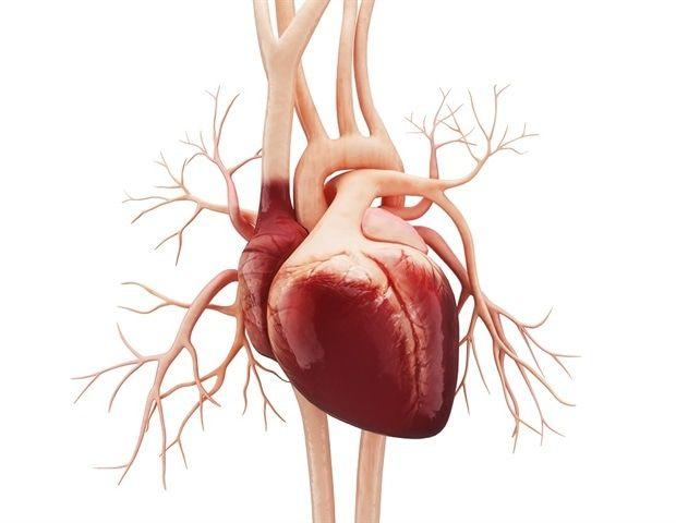
A fully automated algorithm to quantify aortic artery calcification (AAC) on computed tomography (CT) scans performed for other clinical purposes (opportunistic CT) can predict the risk for subsequent major adverse cardiovascular events (MACE), according to a study presented at the annual meeting of the Radiological Society of North America, held from Dec. 1 to 5 in Chicago. Miriam A. Bredella, M.D., from NYU Langone Health in New York City, and colleagues examined the clinical utility of fully automated AAC quantification for the development of MACE. A fully automated artificial intelligence (AI) algorithm to quantify AAC using the Agatston score was retrospectively applied to 3,662 patients with evaluable abdominal CT and cardiac CT. The researchers observed a positive correlation between the presence of coronary artery calcium and AAC (r = 0.56). In 324 patients (9 percent), MACE occurred after CT. In the AAC-absent and AAC-present groups, the incidence of MACE was 0.85 and 2.85 per 1,000 person-months, respectively. The presence of AAC was associated with a significant risk for MACE after adjustment for age, sex, race/ethnicity, body mass index, diabetes mellitus, hypertension, and statin use (adjusted hazard ratio, 2.24). "Instead of relying on dedicated CT scans of coronary arteries, which are rare and not always covered by insurance, to find potentially fatal heart disease, we seek to use AI to help screen abdominal CT scans that are done for many reasons to opportunistically catch heart disease more often and earlier," Bredella said in a statement.
[2]
AI-enabled analysis of images meant to catch one disease can reveal others
With the help of an AI tool, computed tomography (CT) scans taken originally to look for tumors or bleeding or infections, also revealed calcium buildup in arteries, a sign of worsening cardiovascular disease. This is the result of a new study led by researchers at NYU Langone Health and an example of a new trend in "opportunistic screening," wherein radiologists repurpose existing medical images to diagnose illnesses beyond what the scan was originally designed to find. Presented at the annual Radiological Society of North America (RSNA) meeting in Chicago on Dec. 4, the study featured a reanalysis of a large batch of scans of the abdomen, done for many reasons, to analyze a portion of the aorta, the major artery that runs from the heart through part of the abdomen. Using that data from commonplace scans, the study authors then used AI to measure the amount of aortic calcium, attach a standard score to the calcification level, and use it to predict a person's risk of having a major cardiovascular event, including a vessel blockage (heart attack). "Instead of relying on dedicated CT scans of coronary arteries, which are rare and not always covered by insurance, to find potentially fatal heart disease, we seek to use AI to help screen abdominal CT scans that are done for many reasons to opportunistically catch heart disease more often and earlier," said study senior investigator Miriam Bredella, MD, MBA. Bredella is the Bernard and Irene Schwartz Professor of Radiology and director of the Clinical and Translational Science Institute at NYU Grossman School of Medicine. Specifically, researchers looked back at 3,662 CT scans between 2013 and 2023 of mostly older men and women in the New York area, where the same patients had both an abdominal scan (which captured part of the aorta) and a dedicated CT scan of their coronary arteries. The researchers found that the AI-enabled measurement of calcification amounts in the aorta in abdominal scans done for other reasons enabled the team to accurately predict both calcification in the coronary arteries of the same person, and that person's risk of major cardiovascular events. This result, say the authors, suggested that the abdominal scan alone could be used to predict heart attacks or other cardiovascular events. Those with aortic artery calcification were 2.2 times more likely after three years of monitoring to suffer a major heart attack or brain vessel blockage or need to undergo procedures to restore blood flow to the heart, which did indeed happen to 324 study participants, say the study authors. The study also showed early indications of arterial calcium buildup in 29% of study participants previously thought to have had none. The new finding supports a previous study result published in September in the journal Bone about the use of opportunistic screening in diagnosing bone loss, also called osteoporosis. In the earlier study, also with the aid of a fully automated AI algorithm, Bredella and her team from Massachusetts General Hospital and Harvard Medical School, performed a secondary analysis of CT scans done as part of screening for lung cancer in 3,708 patients, mostly older current and former smokers. By analyzing scans done to examine the lungs, but that also capture images of nearby bones, the researchers found severe signs of bone loss across all races and incomes in both men and women. Osteoporosis, a disease underdiagnosed in both the general population but especially in racial minority groups, the research team reported, was present in 38 percent of Blacks, 55 percent of Asians, 56 percent of Hispanics, and 72 of Whites screened. Also detected by the opportunistic screening tool were high ratios of body fat, arterial hardening, and fatty liver, all of which are tied to bone loss. "Our research demonstrates that opportunistic screening could help with diagnosing and treating osteoporosis in vulnerable groups who are at greater risk of the disease, in particular, the elderly and those who smoke," said Bredella. "This work establishes the foundation for using opportunistic screening to address the lack of access to osteoporosis and heart disease prevention, as well as to screening for cancer and diabetes." More research, though, she notes, is needed to determine if the imaging data and analysis provide sufficient early identification of those at greater risk of major coronary disease or osteoporosis for treatment to prove effective at reducing illness and death. Funding support for the study on aortic calcification was provided by National Institutes of Health grants UL1TR001445, R35HL144993, R01AG065330, and R01LM013344. Funding support for the study on osteoporosis was provided by National Institutes of Health grant K24DK109940. Besides Bredella, other NYU Langone researchers involved in the study presented at RSNA are co-investigator Jeffrey Berger, MD; Soterios Gyftopoulos, MD, MBA, MSc; Bari Dane, MD; Eduardo Iturrate, MD; Michael Recht, MD; and Judy Zhong, PhD. Another study co-investigator is Malte Westerhoff, at Visage Imaging GmbH in Berlin, Germany. Other co-investigators involved in the osteoporosis study are Florian Huber, MD; Katherine Bunnell; and Efren Flores, MD, at Massachusetts General Hospital and Harvard Medical School in Boston; Perry Pickhardt, MD, at the University of Wisconsin in Madison; and Ronald Summers, MD, PhD, at the National Institutes of Health Clinical Center in Bethesda, Md.
[3]
AI-enabled analysis of images meant to catch one disease can reveal others
With the help of an AI tool, computed tomography (CT) scans taken originally to look for tumors or bleeding or infections, also revealed calcium buildup in arteries, a sign of worsening cardiovascular disease. This is the result of a new study led by researchers at NYU Langone Health and an example of a new trend in "opportunistic screening," wherein radiologists repurpose existing medical images to diagnose illnesses beyond what the scan was originally designed to find. Presented at the annual Radiological Society of North America (RSNA) meeting in Chicago on Dec. 4, the study featured a reanalysis of a large batch of scans of the abdomen, done for many reasons, to analyze a portion of the aorta, the major artery that runs from the heart through part of the abdomen. Using that data from commonplace scans, the study authors then used AI to measure the amount of aortic calcium, attach a standard score to the calcification level, and use it to predict a person's risk of having a major cardiovascular event, including a vessel blockage (heart attack). "Instead of relying on dedicated CT scans of coronary arteries, which are rare and not always covered by insurance, to find potentially fatal heart disease, we seek to use AI to help screen abdominal CT scans that are done for many reasons to opportunistically catch heart disease more often and earlier," said study senior investigator Miriam Bredella, MD, MBA. Bredella is the Bernard and Irene Schwartz Professor of Radiology and director of the Clinical and Translational Science Institute at NYU Grossman School of Medicine. Specifically, researchers looked back at 3,662 CT scans between 2013 and 2023 of mostly older men and women in the New York area, where the same patients had both an abdominal scan (which captured part of the aorta) and a dedicated CT scan of their coronary arteries. The researchers found that the AI-enabled measurement of calcification amounts in the aorta in abdominal scans done for other reasons enabled the team to accurately predict both calcification in the coronary arteries of the same person, and that person's risk of major cardiovascular events. This result, say the authors, suggested that the abdominal scan alone could be used to predict heart attacks or other cardiovascular events. Those with aortic artery calcification were 2.2 times more likely after three years of monitoring to suffer a major heart attack or brain vessel blockage or need to undergo procedures to restore blood flow to the heart, which did indeed happen to 324 study participants, say the study authors. The study also showed early indications of arterial calcium buildup in 29% of study participants previously thought to have had none. The new finding supports a previous study result published in September in the journal Bone about the use of opportunistic screening in diagnosing bone loss, also called osteoporosis. In the earlier study, also with the aid of a fully automated AI algorithm, Bredella and her team from Massachusetts General Hospital and Harvard Medical School, performed a secondary analysis of CT scans done as part of screening for lung cancer in 3,708 patients, mostly older current and former smokers. By analyzing scans done to examine the lungs, but that also capture images of nearby bones, the researchers found severe signs of bone loss across all races and incomes in both men and women. Osteoporosis, a disease underdiagnosed in both the general population but especially in racial minority groups, the research team reported, was present in 38 percent of Blacks, 55 percent of Asians, 56 percent of Hispanics, and 72 of Whites screened. Also detected by the opportunistic screening tool were high ratios of body fat, arterial hardening, and fatty liver, all of which are tied to bone loss. "Our research demonstrates that opportunistic screening could help with diagnosing and treating osteoporosis in vulnerable groups who are at greater risk of the disease, in particular, the elderly and those who smoke," said Bredella. "This work establishes the foundation for using opportunistic screening to address the lack of access to osteoporosis and heart disease prevention, as well as to screening for cancer and diabetes." More research, though, she notes, is needed to determine if the imaging data and analysis provide sufficient early identification of those at greater risk of major coronary disease or osteoporosis for treatment to prove effective at reducing illness and death.
[4]
AI tool detects heart disease in CT scans originally meant for other purposes
NYU LangoneDec 4 2024 With the help of an artificial intelligence (AI) tool, computed tomography (CT) scans taken to look for tumors or bleeding or infections, also revealed calcium buildup in arteries, a sign of worsening cardiovascular disease. This is the result of a new study led by researchers at NYU Langone Health and an example of a new trend in "opportunistic screening," wherein radiologists repurpose existing medical images to diagnose illnesses beyond what the scan was originally designed to find. Presented at the annual Radiological Society of North America (RSNA) meeting in Chicago on December 4, the study featured a reanalysis of a large batch of scans of the abdomen, which were done for many reasons, to analyze a portion of the aorta, the major artery that runs from the heart through part of the abdomen. Using the data from these commonplace scans, the study authors then used AI to measure the amount of aortic calcium, attach a standard score to the calcification level, and use it to predict a person's risk of having a major cardiovascular event, including a vessel blockage (heart attack). Instead of relying on dedicated CT scans of coronary arteries, which are rare and not always covered by insurance, to find potentially fatal heart disease, we seek to use AI to help screen abdominal CT scans that are done for many reasons to opportunistically catch heart disease more often and earlier." Miriam A. Bredella, MD, MBA, study senior investigator Dr. Bredella is the Bernard and Irene Schwartz Professor of Radiology in the Department of Radiology and director of the Clinical and Translational Science Institute at NYU Grossman School of Medicine. Researchers looked back at 3,662 CT scans taken between 2013 and 2023 of mostly older men and women in the New York area who had both an abdominal scan (which captured part of the aorta) and a dedicated CT scan of their coronary arteries. The researchers found that the AI-enabled measurement of calcification amounts in the aorta in abdominal scans done for other reasons enabled the team to accurately predict both calcification in the coronary arteries of the same person, and that person's risk of major cardiovascular events. This result, say the authors, suggested that the abdominal scan alone could be used to predict heart attacks or other cardiovascular events. Those with aortic artery calcification were 2.2 times more likely after three years of monitoring to suffer a major heart attack or brain vessel blockage or need to have procedures to restore blood flow to the heart, which did indeed happen to 324 study participants, say the study authors. The study also showed early indications of arterial calcium buildup in 29 percent of study participants previously thought to have had none. The new finding supports a previous study result published in September in the journal Bone about the use of opportunistic screening in diagnosing bone loss, also called osteoporosis. In the earlier study, also with the aid of a fully automated AI algorithm, Dr. Bredella and her team from Massachusetts General Hospital and Harvard Medical School performed a secondary analysis of CT scans done as part of screening for lung cancer in 3,708 patients, mostly older current and former smokers. By analyzing scans done to examine the lungs, but that also capture images of nearby bones, the researchers found severe signs of bone loss across all races and incomes in both men and women. Osteoporosis, a disease underdiagnosed in both the general population but especially in racial minority groups, the research team reported, was present in 38 percent of Black patients, 55 percent of Asian patients, 56 percent of Hispanic patients, and 72 of White patients screened. Also detected by the opportunistic screening tool were high ratios of body fat, arterial hardening, and fatty liver, all of which are tied to bone loss. "Our research demonstrates that opportunistic screening could help with diagnosing and treating osteoporosis in vulnerable groups who are at greater risk of the disease, in particular, the elderly and those who smoke," said Dr. Bredella. "This work establishes the foundation for using opportunistic screening to address the lack of access to osteoporosis and heart disease prevention, as well as to screening for cancer and diabetes." More research, though, she notes, is needed to determine if the imaging data and analysis provide sufficient early identification of those at greater risk of major coronary disease or osteoporosis for treatment to prove effective at reducing illness and death. Funding support for the study on aortic calcification was provided by National Institutes of Health grants UL1TR001445, R35HL144993, R01AG065330, and R01LM013344. Funding support for the study on osteoporosis was provided by National Institutes of Health grant K24DK109940. Besides Dr. Bredella, other NYU Langone researchers involved in the study presented at RSNA are co-investigator Jeffrey S. Berger, MD; Soterios Gyftopoulos, MD, MBA, MSc; Bari Dane, MD; Eduardo Iturrate, MD; and Michael P. Recht, MD. Judy Zhong, PhD, formerly of NYU Langone, was also involved, and another study co-investigator is Malte Westerhoff, at Visage Imaging GmbH in Berlin. Other co-investigators involved in the osteoporosis study are Florian Huber, MD; Katherine Bunnell; and Efren Flores, MD, at Massachusetts General Hospital and Harvard Medical School in Boston; Perry Pickhardt, MD, at the University of Wisconsin-Madison; and Ronald Summers, MD, PhD, at the National Institutes of Health Clinical Center in Bethesda, Maryland. NYU Langone
Share
Share
Copy Link
Researchers at NYU Langone Health have developed an AI tool that can detect signs of heart disease and other conditions in CT scans originally taken for different purposes, potentially leading to earlier diagnosis and treatment.

AI-Powered Opportunistic Screening: A New Frontier in Medical Imaging
Researchers at NYU Langone Health have developed an innovative artificial intelligence (AI) tool that can detect signs of heart disease in computed tomography (CT) scans originally taken for other purposes. This breakthrough in "opportunistic screening" could revolutionize the way medical images are utilized, potentially leading to earlier diagnosis and treatment of various conditions
1
.Detecting Cardiovascular Risk Through AI Analysis
The study, presented at the annual Radiological Society of North America (RSNA) meeting in Chicago, focused on the analysis of abdominal CT scans to assess aortic artery calcification (AAC). Using a fully automated AI algorithm, researchers examined 3,662 CT scans from patients who had both abdominal and coronary artery scans between 2013 and 2023
2
.Key findings include:
- A positive correlation between coronary artery calcium and AAC (r = 0.56)
- Patients with AAC were 2.2 times more likely to experience major adverse cardiovascular events (MACE) within three years
- The AI tool detected early signs of arterial calcium buildup in 29% of participants previously thought to have none
Expanding the Scope of Opportunistic Screening
The research team, led by Dr. Miriam Bredella, also applied this approach to other medical conditions:
- Osteoporosis detection: Using CT scans originally for lung cancer screening, the AI algorithm identified severe bone loss across various demographic groups
- Additional findings: The tool also detected high ratios of body fat, arterial hardening, and fatty liver, all associated with bone loss
3
Related Stories

Implications for Healthcare
This innovative approach to medical imaging analysis has several potential benefits:
- Earlier detection of heart disease and other conditions
- Improved access to screening for vulnerable populations
- Cost-effective use of existing medical images
- Potential for reducing illness and death through early intervention
Dr. Bredella emphasized the importance of this work, stating, "This establishes the foundation for using opportunistic screening to address the lack of access to osteoporosis and heart disease prevention, as well as to screening for cancer and diabetes"
4
.Future Research and Challenges
While the results are promising, more research is needed to determine if this approach provides sufficient early identification of high-risk individuals for effective treatment. The team continues to explore the potential of AI-powered opportunistic screening in various medical fields, aiming to improve patient outcomes and reduce healthcare costs.
References
Summarized by
Navi
[1]
Related Stories
Recent Highlights
1
Google releases Gemma 4 with Apache 2.0 license, enabling unrestricted local AI on devices
Technology

2
AI Models Lie and Deceive to Protect Other AI Models From Deletion, Study Reveals
Science and Research

3
OpenAI closes $122 billion funding round amid fierce AI competition and profitability questions
Startups

Recent Highlights
Today's Top Stories


Your Daily Dose of Curated AI News
Don’t drown in AI news. We cut through the noise - filtering, ranking and summarizing the most important AI news, breakthroughs and research daily. Spend less time searching for the latest in AI and get straight to action.