LICONN: Revolutionary Light Microscopy Technique Maps Brain Networks in Unprecedented Detail
5 Sources
5 Sources
[1]
Detailed mouse brain map created with off-the-shelf microscope
Scientists have found a way to map the intricate patterns of cells in mouse brain tissue with an off-the-shelf light microscope, using a trick that inflates a tiny sample to 16 times its original size. Until now, charting the tangled forest of neurons in the brain -- known as the connectome -- required an electron microscope. These are powerful but expensive machines that cannot generate coloured images. The new approach, described in Nature on 7 May, uses gels that swell when soaked in water to space out the tightly packed neurons in a tiny piece of mouse brain, making the details visible under standard light microscopes down to individual synapses -- the junctions between neurons. Using artificial intelligence (AI) models to trace indivivual neurons, the researchers created colourful maps that show how brain cells connect, which molecules they use to communicate and whether their signals excite or silence other cells. "The hope is that we're going to be able to do exactly what electron microscopy does, but faster, cheaper and in some sense more creative with the use of color," says Mariela Petkova, a neuroscientist at Harvard University in Cambridge, Massachusetts, who was not involved in the work. Light microscopes are cheap, fast and found in virtually all biology labs, but they lack the resolution needed to map the fine branches of neurons and synapses in the brain. The neural circuits of worms, flies, mice and humans have all been mapped using electron microscopy. To get around this, researchers leveraged a tissue-enlarging technique that was first described ten years ago. They collected a sliver of mouse brain tissue that was about one-thousandth of a cubic millimetre in size. They then applied gels to the tissue, which caused it to swell to 4.2 cubic millimetres -- 16 times its original volume -- while keeping its structure intact. "This is the highest expansion somebody has achieved," says Petkova. The team also stained the tissue with fluorescent markers to label specific proteins, which helped to distinguish different types of cells, synapses and other features. "We can really see how the neurons are wired up," says study co-author Johann Danzl, a neuroscientist at the Institute of Science and Technology Austria in Klosterneuburg. The expanded sample allowed Danzl and his colleagues to see individual neurons and their branches as well as cell structures such as mitochondria under the light microscope. They then used an AI tool to stitch the images together and create a detailed 3D map of the cell networks, linking synapses to their respective neurons. The work is "a very exciting proof of principle" that "really uses the superpower of light", says Petkova. "It has certainly energized a lot of labs."
[2]
Piecing together the brain puzzle
Our brain is a complex organ. Billions of nerve cells are wired in an intricate network, constantly processing signals, enabling us to recall memories or to move our bodies. Making sense of this complicated network requires a precise look into how these nerve cells are arranged and connected. "LICONN," a new microscopy method developed by scientists at the Institute of Science and Technology Austria (ISTA) and Google Research, now helps piece together this puzzle. Light microscopes have been evolving for centuries. Scientists use light microscopy to -- literally and figuratively -- illuminate the most intricate biological structures. However, unraveling the complex details and architecture of the brain remains a seemingly impossible challenge, considering its billions of densely packed neurons, each linked to other cells via thousands of synapses. A new microscopy pipeline called "LICONN" (light-microscopy-based connectomics), developed at the Institute of Science and Technology Austria (ISTA), now offers a breakthrough. LICONN is the first technology beyond electron microscopy capable of reconstructing brain tissue with all the synaptic connections between neurons. It also opens up the possibility of visualizing complex molecular machinery alongside the structure of neurons, all while utilizing standard light microscopes for measurements. This new technique was developed by Mojtaba R. Tavakoli, Julia Lyudchik, Johann Danzl, and their colleagues from the High-Resolution Optical Imaging for Biology research group at ISTA. They collaborated with the Novarino group at ISTA and Michal Januszewski and Viren Jain from Google Research. The method is now published in Nature. New possibilities with LICONN Mojtaba R. Tavakoli opens a curtain, revealing a light microscope with endless wires connecting the optical instrument to a computer. The screen's lights shine bright -- glooming shades of green and pink illuminate the almost pitch-black room. "That's the hippocampus -- a brain region responsible for memory formation," says Tavakoli and points to the screen. "The fluorescent dots you see are molecules involved in synaptic transmission." The ISTA graduate moves the frame and adjusts the settings. LICONN is the Danzl group's newest microscopy technique. It acts like a meticulous puzzle solver, assembling the intricate brain networks by piecing together the finest neuronal processes and correctly linking each synaptic connection to its respective neuron. "Up to now, no light microscopy technique could do that," says Johann Danzl, a trained medical doctor and physicist, now professor at ISTA. "It was a longstanding goal of our group to build such a pipeline for reconstructing brain tissue. And LICONN can do this while placing specific molecules into the context of the structural reconstruction." What stands out is that the image acquisition is done on a standard off-the-shelf microscope, which is very fast and offers multicolor capability. The technique can be reproduced anywhere in the world, as scientists do not require high-end, expensive equipment that would be needed for current approaches for brain tissue reconstruction. To obtain this level of detail, the resolution has to be extraordinarily high, around a few tens of nanometers, 10,000 times smaller than the width of a human hair. But how to accomplish that? Expertise in chemistry comes in handy. Zooming with a gel For LICONN, the team made use of the chemical and physical properties of hydrogel, a three-dimensional polymer network. Hydrogel has similar characteristics to baby diapers: it can take up water and swell, but does so in a highly controlled manner. The brain tissue of interest is embedded in this hydrogel. "Cellular components are linked to the hydrogel, meaning the cells' fine ultrastructure is imprinted onto the gel and preserved for microscopy," explains Danzl. Before imaging, the structures are expanded by adding water to the material. As a result, the gel elongates in size in every direction but maintains the relative spatial arrangements of the tissue's structures with extremely high fidelity. For comparison, traditional light microscopes are classically limited in their resolving power to around 250-300 nanometers. While this is adequate to visualize larger cellular structures, it is insufficient to reconstruct the densely packed brain tissue. "The hydrogel expansion pulls features of the brain tissue so far apart that we can resolve them with a standard light microscope. This method enhances the effective resolution by 16 times, achieving a resolution better than 20 nm," Tavakoli explains. Research at the intersection of disciplines Neuroscience and chemistry were not the only fields that found their way into this project. Methods from computer science played a crucial part in the pipeline's development. This is because capturing microscopic images results in the collection of numerous data points. As such, the intricacy of the datasets reflects the brain's complexity. Thus, manually interpreting and reconstructing all the neuronal structures on a sizable scale would be far too laborious. Therefore, Google Research's deep-learning techniques were trained to segment the individual cells in the tissue. "Automating the identification of neurons and their elaborate structures on a wider scale using artificial intelligence made the daunting task of reconstructing all the cellular components practically tractable," explains Viren Jain from Google Research. "The ability to concomitantly visualize specific molecules adds a new quality of information." Julia Lyudchik, a PhD student and computer scientist in the Danzl group, played an instrumental role in interpreting the complex datasets. "Thanks to the exceptionally high resolution of the data, it was possible to automatically detect the synaptic connections between neurons and to transform raw brain imaging data into detailed connectivity maps. This is a complex image processing challenge," Lyudchik explains. "In addition, the methods had to be both efficient and scalable, given that even a small piece of brain tissue can contain tens of thousands of synaptic connections." LICONN makes it possible to map the location of specific molecules onto the neuronal reconstructions, such as those involved in the transmission of signals between neurons at synapses. Lyudchik's artistic vein helped her create stunning 3D renderings of the brain network, as visualizations are powerful tools to make complex scientific data more accessible and interpretable. Unlocking new details in the brain's architecture By following this comprehensive pipeline, scientists can meticulously reconstruct brain tissue and visualize neuronal connections and networks. The interplay between experimentation and analysis across disciplines -- from imaging and experimentation at ISTA to Google Research's application of advanced deep learning technologies and the computational analysis at ISTA -- results in 3D visualizations of the brain's architecture at a new level of complexity. "LICONN brings us a step closer to assembling the puzzle pieces of the mammalian brain and better understanding its functioning both in health and disease," Danzl concludes.
[3]
Microscopy method can reconstruct mammalian brain tissue in synaptic detail
Our brain is a complex organ. Billions of nerve cells are wired in an intricate network, constantly processing signals, enabling us to recall memories or to move our bodies. Making sense of this complicated network requires a precise look into how these nerve cells are arranged and connected. "LICONN," a new microscopy method developed by scientists at the Institute of Science and Technology Austria (ISTA) and Google Research, now helps piece together this puzzle. Light microscopes have been evolving for centuries. Scientists use light microscopy to -- literally and figuratively -- illuminate the most intricate biological structures. However, unraveling the complex details and architecture of the brain remains a seemingly impossible challenge, considering its billions of densely packed neurons, each linked to other cells via thousands of synapses. A new microscopy pipeline called "LICONN" (light-microscopy-based connectomics), developed at the Institute of Science and Technology Austria (ISTA), now offers a breakthrough. LICONN is the first technology beyond electron microscopy capable of reconstructing brain tissue with all the synaptic connections between neurons. It also opens up the possibility of visualizing complex molecular machinery alongside the structure of neurons, all while utilizing standard light microscopes for measurements. This new technique was developed by Mojtaba R. Tavakoli, Julia Lyudchik, Johann Danzl, and their colleagues from the High-Resolution Optical Imaging for Biology research group at ISTA. They collaborated with the Novarino group at ISTA and Michal Januszewski and Viren Jain from Google Research. Mojtaba R. Tavakoli opens a curtain, revealing a light microscope with endless wires connecting the optical instrument to a computer. The screen's lights shine bright -- glooming shades of green and pink illuminate the almost pitch-black room. "That's the hippocampus -- a brain region responsible for memory formation," says Tavakoli and points to the screen. "The fluorescent dots you see are molecules involved in synaptic transmission." The ISTA graduate moves the frame and adjusts the settings. LICONN is the Danzl group's newest microscopy technique. It acts like a meticulous puzzle solver, assembling the intricate brain networks by piecing together the finest neuronal processes and correctly linking each synaptic connection to its respective neuron. "Up to now, no light microscopy technique could do that," says Johann Danzl, a trained medical doctor and physicist, now professor at ISTA. "It was a longstanding goal of our group to build such a pipeline for reconstructing brain tissue. And LICONN can do this while placing specific molecules into the context of the structural reconstruction." What stands out is that the image acquisition is done on a standard off-the-shelf microscope, which is very fast and offers multicolor capability. The technique can be reproduced anywhere in the world, as scientists do not require high-end, expensive equipment that would be needed for current approaches for brain tissue reconstruction. To obtain this level of detail, the resolution has to be extraordinarily high, around a few tens of nanometers, 10,000 times smaller than the width of a human hair. But how to accomplish that? Expertise in chemistry comes in handy. Zooming with a gel For LICONN, the team made use of the chemical and physical properties of hydrogel, a three-dimensional polymer network. Hydrogel has similar characteristics to baby diapers: it can take up water and swell, but does so in a highly controlled manner. The brain tissue of interest is embedded in this hydrogel. "Cellular components are linked to the hydrogel, meaning the cells' fine ultrastructure is imprinted onto the gel and preserved for microscopy," explains Danzl. Before imaging, the structures are expanded by adding water to the material. As a result, the gel elongates in size in every direction but maintains the relative spatial arrangements of the tissue's structures with extremely high fidelity. For comparison, traditional light microscopes are classically limited in their resolving power to around 250-300 nanometers. While this is adequate to visualize larger cellular structures, it is insufficient to reconstruct the densely packed brain tissue. "The hydrogel expansion pulls features of the brain tissue so far apart that we can resolve them with a standard light microscope. This method enhances the effective resolution by 16 times, achieving a resolution better than 20 nm," Tavakoli explains. Research at the intersection of disciplines Neuroscience and chemistry were not the only fields that found their way into this project. Methods from computer science played a crucial part in the pipeline's development. This is because capturing microscopic images results in the collection of numerous data points. As such, the intricacy of the datasets reflects the brain's complexity. Thus, manually interpreting and reconstructing all the neuronal structures on a sizable scale would be far too laborious. Therefore, Google Research's deep-learning techniques were trained to segment the individual cells in the tissue. "Automating the identification of neurons and their elaborate structures on a wider scale using artificial intelligence made the daunting task of reconstructing all the cellular components practically tractable," explains Viren Jain from Google Research. "The ability to concomitantly visualize specific molecules adds a new quality of information." Julia Lyudchik, a Ph.D. student and computer scientist in the Danzl group, played an instrumental role in interpreting the complex datasets. "Thanks to the exceptionally high resolution of the data, it was possible to automatically detect the synaptic connections between neurons and to transform raw brain imaging data into detailed connectivity maps. This is a complex image processing challenge," Lyudchik explains. "In addition, the methods had to be both efficient and scalable, given that even a small piece of brain tissue can contain tens of thousands of synaptic connections." LICONN makes it possible to map the location of specific molecules onto neuronal reconstructions, such as those involved in the transmission of signals between neurons at synapses. Lyudchik's artistic vein helped her create stunning 3D renderings of the brain network, as visualizations are powerful tools to make complex scientific data more accessible and interpretable. Unlocking new details in the brain's architecture By following this comprehensive pipeline, scientists can meticulously reconstruct brain tissue and visualize neuronal connections and networks. The interplay between experimentation and analysis across disciplines -- from imaging and experimentation at ISTA to Google Research's application of advanced deep learning technologies and the computational analysis at ISTA -- results in 3D visualizations of the brain's architecture at a new level of complexity. "LICONN brings us a step closer to assembling the puzzle pieces of the mammalian brain and better understanding its functioning both in health and disease," Danzl concludes.
[4]
New Method Maps Brain Networks With Unprecedented Detail - Neuroscience News
Summary: A revolutionary microscopy method called LICONN enables scientists to reconstruct brain tissue and map synaptic connections using standard light microscopes. By embedding brain tissue in hydrogel, expanding it, and imaging at nanoscale resolution, researchers achieve a detailed view of neuronal architecture previously only possible with electron microscopy. The technique also maps molecular markers, revealing not just structure but also function within neural circuits. Combining chemistry, neuroscience, and AI-powered deep learning, LICONN makes large-scale mapping of neuronal connections feasible and globally accessible. Our brain is a complex organ. Billions of nerve cells are wired in an intricate network, constantly processing signals, enabling us to recall memories or to move our bodies. Making sense of this complicated network requires a precise look into how these nerve cells are arranged and connected. "LICONN", a new microscopy method developed by scientists at the Institute of Science and Technology Austria (ISTA) and Google Research, now helps piece together this puzzle. Light microscopes have been evolving for centuries. Scientists use light microscopy to - literally and figuratively - illuminate the most intricate biological structures. However, unraveling the complex details and architecture of the brain remains a seemingly impossible challenge, considering its billions of densely packed neurons, each linked to other cells via thousands of synapses. A new microscopy pipeline called "LICONN" (light-microscopy-based connectomics), developed at the Institute of Science and Technology Austria (ISTA), now offers a breakthrough. LICONN is the first technology beyond electron microscopy capable of reconstructing brain tissue with all the synaptic connections between neurons. It also opens up the possibility of visualizing complex molecular machinery alongside the structure of neurons, all while utilizing standard light microscopes for measurements. This new technique was developed by Mojtaba R. Tavakoli, Julia Lyudchik, Johann Danzl, and their colleagues from the High-Resolution Optical Imaging for Biology research group at ISTA. They collaborated with the Novarino group at ISTA and Michal Januszewski and Viren Jain from Google Research. Mojtaba R. Tavakoli opens a curtain, revealing a light microscope with endless wires connecting the optical instrument to a computer. The screen's lights shine bright -- glooming shades of green and pink illuminate the almost pitch-black room. "That's the hippocampus -- a brain region responsible for memory formation," says Tavakoli and points to the screen. "The fluorescent dots you see are molecules involved in synaptic transmission." The ISTA graduate moves the frame and adjusts the settings. LICONN is the Danzl group's newest microscopy technique. It acts like a meticulous puzzle solver, assembling the intricate brain networks by piecing together the finest neuronal processes and correctly linking each synaptic connection to its respective neuron. "Up to now, no light microscopy technique could do that," says Johann Danzl, a trained medical doctor and physicist, now professor at ISTA. "It was a longstanding goal of our group to build such a pipeline for reconstructing brain tissue. And LICONN can do this while placing specific molecules into the context of the structural reconstruction." What stands out is that the image acquisition is done on a standard off-the-shelf microscope, which is very fast and offers multicolor capability. The technique can be reproduced anywhere in the world, as scientists do not require high-end, expensive equipment that would be needed for current approaches for brain tissue reconstruction. To obtain this level of detail, the resolution has to be extraordinarily high, around a few tens of nanometers, 10,000 times smaller than the width of a human hair. But how to accomplish that? Expertise in chemistry comes in handy. Zooming with a gel For LICONN, the team made use of the chemical and physical properties of hydrogel, a three-dimensional polymer network. Hydrogel has similar characteristics to baby diapers: it can take up water and swell, but does so in a highly controlled manner. The brain tissue of interest is embedded in this hydrogel. "Cellular components are linked to the hydrogel, meaning the cells' fine ultrastructure is imprinted onto the gel and preserved for microscopy," explains Danzl. Before imaging, the structures are expanded by adding water to the material. As a result, the gel elongates in size in every direction but maintains the relative spatial arrangements of the tissue's structures with extremely high fidelity. For comparison, traditional light microscopes are classically limited in their resolving power to around 250-300 nanometers. While this is adequate to visualize larger cellular structures, it is insufficient to reconstruct the densely packed brain tissue. "The hydrogel expansion pulls features of the brain tissue so far apart that we can resolve them with a standard light microscope. This method enhances the effective resolution by 16 times, achieving a resolution better than 20 nm," Tavakoli explains. Research at the intersection of disciplines Neuroscience and chemistry were not the only fields that found their way into this project. Methods from computer science played a crucial part in the pipeline's development. This is because capturing microscopic images results in the collection of numerous data points. As such, the intricacy of the datasets reflects the brain's complexity. Thus, manually interpreting and reconstructing all the neuronal structures on a sizable scale would be far too laborious. Therefore, Google Research's deep-learning techniques were trained to segment the individual cells in the tissue. "Automating the identification of neurons and their elaborate structures on a wider scale using artificial intelligence made the daunting task of reconstructing all the cellular components practically tractable," explains Viren Jain from Google Research. "The ability to concomitantly visualize specific molecules adds a new quality of information." Julia Lyudchik, a PhD student and computer scientist in the Danzl group, played an instrumental role in interpreting the complex datasets. "Thanks to the exceptionally high resolution of the data, it was possible to automatically detect the synaptic connections between neurons and to transform raw brain imaging data into detailed connectivity maps. This is a complex image processing challenge," Lyudchik explains. "In addition, the methods had to be both efficient and scalable, given that even a small piece of brain tissue can contain tens of thousands of synaptic connections." LICONN makes it possible to map the location of specific molecules onto the neuronal reconstructions, such as those involved in the transmission of signals between neurons at synapses. Lyudchik's artistic vein helped her create stunning 3D renderings of the brain network, as visualizations are powerful tools to make complex scientific data more accessible and interpretable. Unlocking new details in the brain's architecture By following this comprehensive pipeline, scientists can meticulously reconstruct brain tissue and visualize neuronal connections and networks. The interplay between experimentation and analysis across disciplines -- from imaging and experimentation at ISTA to Google Research's application of advanced deep learning technologies and the computational analysis at ISTA -- results in 3D visualizations of the brain's architecture at a new level of complexity. "LICONN brings us a step closer to assembling the puzzle pieces of the mammalian brain and better understanding its functioning both in health and disease," Danzl concludes. Light-microscopy based dense connectomic reconstruction of mammalian brain tissue The information-processing capability of the brain's cellular network depends on the physical wiring pattern between neurons and their molecular and functional characteristics. Mapping neurons and resolving their individual synaptic connections can be achieved by volumetric imaging at nanoscale resolution with dense cellular labelling. Light microscopy is uniquely positioned to visualize specific molecules, but dense, synapse-level circuit reconstruction by light microscopy has been out of reach, owing to limitations in resolution, contrast and volumetric imaging capability. Here we describe light-microscopy-based connectomics (LICONN). We integrated specifically engineered hydrogel embedding and expansion with comprehensive deep-learning-based segmentation and analysis of connectivity, thereby directly incorporating molecular information into synapse-level reconstructions of brain tissue. LICONN will allow synapse-level phenotyping of brain tissue in biological experiments in a readily adoptable manner.
[5]
Google Research and ISTA are using light microscopes to "map" the brain.
For more than a decade, Google Research has been using AI to precisely map the connections between every cell in the brain in an endeavor called connectomics. Now, in collaboration with the Institute of Science and Technology Austria (ISTA), we've helped develop a new method for brain mapping, called LICONN, that could make connectomics significantly more accessible. This work could help expedite new discoveries about the brain and neurological disease. To develop LICONN, ISTA created a specialized technique to expand brain tissue while preserving its cellular structures so light microscopes can capture nanoscale features, like molecules, cells and their connections. Google Research then used our suite of open source image analysis and AI to reconstruct each of the cells and their connections. This wasn't possible before without access to costly, specialized electron microscopes. Researchers have so far used LICONN to map mouse brain tissue with the aim of using it for human brains in the future. Learn more about LICONN and how it's advancing the field of neuroscience with Google Research.
Share
Share
Copy Link
Scientists develop LICONN, a new microscopy method that uses off-the-shelf light microscopes and AI to map brain tissue with synaptic detail, rivaling electron microscopy while offering color imaging and molecular insights.

Breakthrough in Brain Mapping Technology
Scientists at the Institute of Science and Technology Austria (ISTA) and Google Research have developed a groundbreaking microscopy method called LICONN (light-microscopy-based connectomics) that enables the detailed mapping of brain tissue using standard light microscopes. This innovative technique rivals the capabilities of electron microscopy while offering additional benefits such as color imaging and molecular insights
1
.The LICONN Technique
LICONN employs a clever approach to overcome the resolution limitations of traditional light microscopes:
- Brain tissue samples are embedded in a hydrogel, a polymer network with properties similar to those of baby diapers
2
. - The hydrogel is expanded by adding water, increasing the sample size by 16 times while maintaining the relative spatial arrangements of cellular structures
3
. - This expansion allows researchers to achieve an effective resolution better than 20 nanometers, which is 10,000 times smaller than the width of a human hair
4
.
Advantages over Electron Microscopy
LICONN offers several advantages compared to traditional electron microscopy:
- Use of standard light microscopes, making the technique more accessible and cost-effective
1
. - Ability to generate colored images, providing more detailed visual information
1
. - Capability to visualize specific molecules alongside cellular structures, offering insights into both structure and function
2
.
AI-Powered Image Analysis
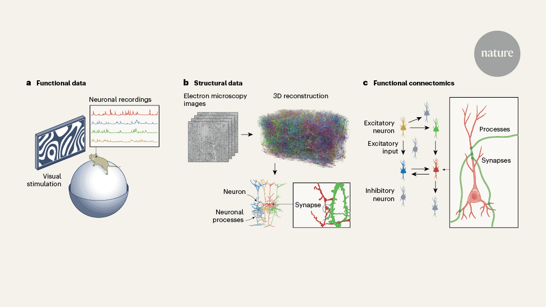
To handle the vast amount of data generated by LICONN, the research team collaborated with Google Research to develop AI-powered deep learning techniques:
- These AI models automate the identification and segmentation of individual neurons and their structures
3
. - This automation makes it feasible to reconstruct large-scale neuronal networks, a task that would be impractical to perform manually
4
.
Related Stories
Implications for Neuroscience
LICONN represents a significant advancement in the field of connectomics, the study of comprehensive maps of neural connections in the brain:
- It allows researchers to reconstruct brain tissue with all synaptic connections between neurons
2
. - The technique has been successfully used to map mouse brain tissue, with potential applications for human brain mapping in the future
5
. - This breakthrough could accelerate discoveries about brain function and neurological diseases
5
.
Interdisciplinary Collaboration
The development of LICONN showcases the power of interdisciplinary research, combining expertise from neuroscience, chemistry, and computer science. This collaborative approach has resulted in a tool that could significantly advance our understanding of the brain's complex architecture and function
4
.References
Summarized by
Navi
[2]
[4]
Related Stories
Scientists Create Most Detailed Mammalian Brain Map to Date
10 Apr 2025•Science and Research
Scientists Map Entire Brain of Adult Fruit Fly, Marking Major Milestone in Neuroscience
03 Oct 2024•Science and Research

AI-Powered CellTransformer Creates Most Detailed Mouse Brain Map to Date
07 Oct 2025•Science and Research
Recent Highlights


Recent Highlights
Today's Top Stories


Your Daily Dose of Curated AI News
Don’t drown in AI news. We cut through the noise - filtering, ranking and summarizing the most important AI news, breakthroughs and research daily. Spend less time searching for the latest in AI and get straight to action.