Scientists Create Most Detailed Mammalian Brain Map to Date
17 Sources
17 Sources
[1]
A map of neural signals and circuits traces the logic of brain computation
You have full access to this article via Jozef Stefan Institute. Millions of years of evolution have endowed animals with cognitive abilities that can surpass modern artificial intelligence. Machine learning requires extensive data sets for training, whereas a mouse that explores an unfamiliar maze and randomly stumbles upon a reward can remember the location of the prize after a handful of successful journeys. To shine a light on the computational circuitry of the mouse brain, researchers from institutes across the United States have led the collaborative MICrONS (Machine Intelligence from Cortical Networks) project and created the most comprehensive data set ever assembled that links mammalian brain structure to neuronal function in an active animal. What distinguishes the mammalian brain from that of other vertebrates is a region called the neocortex, which is the largest and evolutionarily newest part of the human brain. The researchers focused on this region because it is generally considered to be the seat of higher cognition and plays a key part in sensory perception, language processing, planning and decision-making. Remarkably, these seemingly different functions are made possible by a blueprint that can be found, with some modifications, in all cortical areas and in all mammals. This makes studying the cortex in some ways similar to working out the principles of a combustion engine by looking at many cars -- there are different engine models, but the same fundamental mechanics apply. To understand the engine, it is useful not only to have a description of all its parts, but also to understand how the parts work together. The MICrONS team applied this idea to the brain. Just as an engine is composed of pistons, cylinders and a fuel system, the brain consists of neurons and synapses -- the tiny, specialized connections at which neurons communicate. To reveal these intricate structures, the MICrONS team used a technique called volume electron microscopy, the gold standard for imaging at a nanometre-scale resolution. It captures exceptionally detailed images of synapses and the neurons they belong to, and enables the exact reconstruction of neural circuits. The process involved slicing a portion of the cortex responsible for visual processing into 28,000 extraordinarily thin sections, each only a few tens of nanometres thick -- more than one thousand times thinner than a human hair. The sections were then imaged with an electron beam and digitally reassembled into a 3D reconstruction of the brain volume. Machine-learning algorithms subsequently treated this volume like a 3D colouring book, automatically filling in the shapes of neurons and systematically labelling vast numbers of synapses. This anatomical data set spans a cubic millimetre of mouse cortex, about the size of a small poppy seed. Yet it contains an astonishing 84,000 neurons, half a billion synapses and 5.4 kilometres of neuronal wiring -- nearly one and a half times the length of New York City's Central Park (estimated from ref. 4). Although this volume represents a tiny fraction of the mouse brain (and an even tinier fraction of the human brain), it pushes the technological limits of the field of connectomics, which is dedicated to mapping the brain into comprehensive wiring diagrams known as connectomes. By comparison, landmark connectomes such as those of the roundworm Caenorhabditis elegans (302 neurons, 5,000 synapses), fruit fly larva (Drosophila melanogaster; 3,016 neurons, 548,000 synapses), and the adult fruit fly central brain (25,000 neurons, 20 million synapses) and whole brain (almost 140,000 neurons, 50 million synapses) are a fraction of this volume. An important connectomics study that compared mouse, rhesus macaque (Macaca mulatta) and human cortical architecture analysed about 1.6 million synapses across the data sets, but the only volume of comparable scale to MICrONS is the one cubic millimetre of human cortex (16,000 neurons, 150 million synapses) that was mapped last year. Together, these two projects define the current technological frontier of large-scale mammalian connectomics. By itself, a blueprint might be insufficient for understanding how an engine works. Before the group generated their nanometre-scale parts list, they had already observed the components in action. This is where the study truly breaks new ground and transforms structural connectomics, which maps anatomy, into functional connectomics, linking that wiring with neuronal activity, on a large scale (Fig. 1). The researchers recorded activity in the cortex from many neurons in parallel, using a genetically modified mouse -- the neurons of which produced a fluorescent protein that emitted light when the cells fired. During many sessions over the course of a few days, the mouse viewed an extensive collection of video clips to activate its visual cortex while its head was stabilized under the microscope, but it could still walk on a treadmill. The activity of 76,000 neurons was recorded simultaneously while tracking the mouse's running speed, eye movements and other facial features. This neuronal activity data was then precisely aligned with the electron microscopy volume by matching individual neurons across the functional and electron-microscopy data sets. Of course, the work shares some of the common limitations of connectomics. Because connectomic wiring diagrams are inherently incomplete, owing to errors in machine-learning-based neuron reconstructions, they require extensive human editing, which is a formidable challenge at this scale. Alleviating the human editing task with next-generation machine-learning models that are tailored to recognize complete neuron shapes is fertile ground for technological development, and first attempts at this have already aided the fast mapping of network motifs in the cortex. At the same time, studying the cortex poses its own challenges. Before starting to explain an engine, it helps to understand clearly what purpose it fulfils in a vehicle. Even though scientists know a lot about how visual features are represented in the cortex, it is much harder to grasp the nature of the 'higher' computations that underlie cortical function. The visual cortex, for example, processes more than just visual signals, and this might explain some of the neuronal activity that was unrelated to the movies presented to the mouse. Finally, cortical circuits are vast and extend beyond the cubic millimetre imaged here, which means no circuit in the data set is truly complete. Despite these limitations, this work marks a major leap forwards and offers an invaluable community resource for future discoveries in neuroscience. In a broader context, connectomics is propelling neuroscience into the era of large-scale team science at the intersection of academia and industry, much like the collaborative efforts behind the Large Hadron Collider at CERN, Europe's particle-physics laboratory near Geneva, Switzerland, for particle physics or the Sloan Digital Sky Survey (SDSS) in New Mexico, for astrophysics. These projects demonstrate the power of interdisciplinary, multi-institutional collaborations fuelled by massive data sets. Just as CERN's investigations into fundamental particles and SDSS's cosmic mapping have transformed our understanding of matter and the universe, the investment in MICrONS will help to unravel the complex neural networks underlying cognition and behaviour.
[2]
Biggest brain map ever details huge number of neurons and their activity
Researchers have created the largest and most detailed wiring diagram of a mammalian brain to date, by mapping cells in a cubic millimetre of a mouse's brain tissue. In a landmark achievement, the diagram also details the activity of individual neurons on a large scale -- a neuroscience first. The high-resolution 3D map contains more than 200,000 brain cells, around 82,000 of which are neurons. It also includes more than 500 million of the neuronal connection points called synapses and more than 4 kilometres of neuronal wiring, all found in a tiny block of tissue in a brain region involved in vision. The only brain map of comparable scale is that of a cubic millimetre of human brain, which included 16,000 neurons and 150 million synapses. The new map also captured the activity of tens of thousands of neurons firing signals and interacting with each other to process visual information. This brain-activity map, combined with the wiring diagram, marks a milestone in connectomics, a field that aims to show how brains process and organize information. Behind the massive efforts are more than 150 researchers in the Machine Intelligence from Cortical Networks (MICrONS) project, who described their work in a package of eight papers published today in Nature and Nature Methods. The MICrONS project has made its resources available for the neuroscience community online, and other teams are already exploring them in different studies. "They managed to do something that we haven't done as a neuroscience community in basically all of our history, which is to be able to map the activity of neurons onto the wiring on a very large population of neurons," says Mariela Petkova, a neuroscientist at Harvard University in Cambridge, Massachusetts, who is not involved with the project. "We have never seen it at this scale." The data "are really stunningly beautiful," says Forrest Collman, a neuroscientist at the Allen Institute for Brain Science in Seattle, Washington, who co-authored the studies. "Looking at it really gives you an awe about the sense of complexity in the brain that is very much akin to looking up at the stars of night." To create the breakthrough map, researchers first recorded the firing of almost 76,000 neurons in the visual cortex of a mouse as the animal watched various videos, including clips from The Matrix, for two hours. Then they sliced up a cubic millimetre of the mouse's brain into thousands of tissue slices, each about one four-hundredth the width of a human hair. The scientists imaged each slice and assembled the images into a 3D map. Finally, they used artificial intelligence and machine-learning algorithms to annotate the neurons, their branching projections and their synapses. The team also matched the neurons in the map with their recordings of brain cells in action. Moritz Helmstaedter, a neuroscientist at the Max Planck Institute for Brain Research in Frankfurt, Germany, says "the combination of function and structure at that scale" is unprecedented. It's "a very impressive endeavour and success". The work yielded insights into the basic rules that shape neural circuits in the mouse brain. For example, the authors found that neurons in the cortex that respond to similar types of visual feature -- such as certain shapes or directions of movement -- often form more connections with one another, no matter how far apart they are, than they do with neurons that specialize in a different type of feature. The results add a new wrinkle to a long-held theory in neuroscience, says Collman -- namely, that "neurons that fire together wire together". Previous studies have tested this theory only in limited numbers of neurons and synapses. The current study shows that "there's a diversity [to] how much this rule is applied across all the different components of cortex", he adds. The MICrONS researchers hope that their data set can help to reveal various features and processes in the brain. "There are all sorts of cortical areas that we understand at different levels of detail and in different ways. And I think this is really only the beginning of relating structure and function," says Clay Reid, a neurobiologist at the Allen Institute and a co-author of the MICrONS papers. Helmstaedter says researchers can use the wiring maps to study how the brain stores and recalls visual memories, such as "our recollection of the last birthday party or of our grandparents". These are "the big open questions about the mammalian cortex that are still very fundamental," he adds. The newly published map covers around 0.2% of the mouse's brain, but the MICrONS team will be testing the technologies to map the animal's entire brain, says Nuno Maçarico da Costa, a neuroanatomist at the Allen Institute and another co-author of the MICrONS papers.
[3]
Largest mammalian brain map ever could unpick what makes us human
A map of part of a mouse brain, which is expected to be generalisable to people, could help scientists understand behaviours, consciousness and even what it means to be human The largest and most comprehensive 3D map of a mammalian brain to date offers an unprecedented insight into how neurons connect and function. The new map, which captures a cubic millimetre of a mouse's visual cortex, will allow scientists to study brain function in extraordinary detail, potentially revealing crucial insights into how neural activity shapes behaviour, how complex traits like consciousness arise, and even what it means to be human. "Our behaviours ultimately arise from activity in the brain, and brain tissue shares very similar properties in all mammals," says team member Forrest Collman at the Allen Institute for Brain Science in Seattle. "This is one reason we believe insights about the mouse cortex can generalise to humans." The achievement - something that biologist Francis Crick said in 1979 was "impossible" - took seven years to complete and involved 150 researchers from three institutions. It began with a team recording neural activity from a portion of a mouse's visual cortex, that was no bigger than a grain of sand, as it watched movies and YouTube clips. Next, a second group dissected that same brain region, dividing it into layers 1/400th the width of a human hair, and took pictures of each slice. Due to the delicate nature of the structure, the slicing process couldn't be stopped for long, so the team took shifts. "We spent 12 days and 12 nights sectioning this millimetre cube of tissue into almost 30,000 layers," says team member Nuno da Costa, also at the Allen Institute. From there, a third team used AI to trace all the cells and reconstruct each slice into a 3D map. "It was like asking AI to do the world's hardest colouring book," says Collman. "You have 100 million images in three dimensions and every single cell has to get coloured with a different crayon. The AI has to decide where one cell starts and the next one stops." This data was finally combined with the functional activity recorded at the start of the project so that what the mouse was watching could be linked with the corresponding activity in the brain. The resulting map illustrates the staggering complexity of the brain. Despite its diminutive size, it contained more than 200,000 cells with 4 kilometres of branches between them, and 523 million synapses joining the cells together. The data is already challenging assumptions about how neurons communicate, revealing that they not only target nearby cells, but actively hunt out other cells dedicated to processing the same visual stimuli. The researchers hope their map will bridge some of the gaps in knowledge between neural activity and behaviours, eventually helping unravel complex traits like intelligence. "It is ground-breaking work that will be invaluable to the scientific community," says Nathalie Rochefort at the University of Edinburgh, UK. Beyond its immediate applications, da Costa says we might even be able to test theories of consciousness. "If someone has a theory of consciousness, they might be able to ask questions of this data, which could then support their theory or reject it." The work builds on another study published last year that mapped every neuron in the adult fly brain - a breakthrough that has already revolutionised the field, says Rochefort. For instance, it has helped scientists better understand the circadian rhythms that affect everything from sleep to metabolism. She says this new map will be invaluable, allowing researchers to make comparisons between it and other maps of different species to examine what cells, wiring principles and functional properties are specific to one species or conserved across several of them, "ultimately shedding light on what makes us human".
[4]
Scientists map part of a mouse's brain that's so complex it looks like a galaxy
WASHINGTON (AP) -- Thanks to a mouse watching clips from "The Matrix," scientists have created the largest functional map of a brain to date - a diagram of the wiring connecting 84,000 neurons as they fire off messages. Using a piece of that mouse's brain about the size of a poppy seed, the researchers identified those neurons and traced how they communicated via branch-like fibers through a surprising 500 million junctions called synapses. The massive dataset, published Wednesday by the journal Nature, marks a step toward unraveling the mystery of how our brains work. The data, assembled in a 3D reconstruction colored to delineate different brain circuitry, is open to scientists worldwide for additional research - and for the simply curious to take a peek. "It definitely inspires a sense of awe, just like looking at pictures of the galaxies," said Forrest Collman of the Allen Institute for Brain Science in Seattle, one of the project's leading researchers. "You get a sense of how complicated you are. We're looking at one tiny part ... of a mouse's brain and the beauty and complexity that you can see in these actual neurons and the hundreds of millions of connections between them." How we think, feel, see, talk and move are due to neurons, or nerve cells, in the brain - how they're activated and send messages to each other. Scientists have long known those signals move from one neuron along fibers called axons and dendrites, using synapses to jump to the next neuron. But there's less known about the networks of neurons that perform certain tasks and how disruptions of that wiring could play a role in Alzheimer's, autism or other disorders. "You can make a thousand hypotheses about how brain cells might do their job but you can't test those hypotheses unless you know perhaps the most fundamental thing - how are those cells wired together," said Allen Institute scientist Clay Reid, who helped pioneer electron microscopy to study neural connections. With the new project, a global team of more than 150 researchers mapped neural connections that Collman compares to tangled pieces of spaghetti winding through part of the mouse brain responsible for vision. The first step: Show a mouse video snippets of sci-fi movies, sports, animation and nature. A team at Baylor College of Medicine did just that, using a mouse engineered with a gene that makes its neurons glow when they're active. The researchers used a laser-powered microscope to record how individual cells in the animal's visual cortex lit up as they processed the images flashing by. Next, scientists at the Allen Institute analyzed that small piece of brain tissue, using a special tool to shave it into more than 25,000 layers, each far thinner than a human hair. With electron microscopes, they took nearly 100 million high-resolution images of those sections, illuminating those spaghetti-like fibers and painstakingly reassembling the data in 3D. Finally, Princeton University scientists used artificial intelligence to trace all that wiring and "paint each of the individual wires a different color so that we can identify them individually," Collman explained. They estimated that microscopic wiring, if laid out, would measure more than 3 miles (5 kilometers). Importantly, matching up all that anatomy with the activity in the mouse's brain as it watched movies allowed researchers to trace how the circuitry worked. The Princeton researchers also created digital 3D copies of the data that other scientists can use in developing new studies. Could this kind of mapping help scientists eventually find treatments for brain diseases? The researchers call it a foundational step, like how the Human Genome Project that provided the first gene mapping eventually led to gene-based treatments. Mapping a full mouse brain is one next goal. "The technologies developed by this project will give us our first chance to really identify some kind of abnormal pattern of connectivity that gives rise to a disorder," another of the project's leading researchers, Princeton neuroscientist and computer scientist Sebastian Seung, said in a statement. The work "marks a major leap forwards and offers an invaluable community resource for future discoveries," wrote Harvard neuroscientists Mariela Petkova and Gregor Schuhknecht, who weren't involved in the project. The huge and publicly shared data "will help to unravel the complex neural networks underlying cognition and behavior," they added. The Machine Intelligence from Cortical Networks, or MICrONS, consortium was funded by the National Institutes of Health's BRAIN Initiative and IARPA, the Intelligence Advanced Research Projects Activity. -- - The Associated Press Health and Science Department receives support from the Howard Hughes Medical Institute's Science and Educational Media Group and the Robert Wood Johnson Foundation. The AP is solely responsible for all content.
[5]
World's Most Detailed Brain Map Built From a Grain of Brain Tissue - Neuroscience News
Summary: Scientists have created the most detailed wiring diagram of a mammalian brain to date, mapping every cell and synapse in a cubic millimeter of a mouse's visual cortex. Using cutting-edge microscopy, AI, and 3D reconstruction, researchers captured more than 200,000 cells and over 500 million connections. The work revealed surprising principles of brain organization, including new inhibitory cell behaviors and network-wide coordination. This achievement provides a foundational tool for understanding brain function, intelligence, and neurological disorders. From a tiny sample of tissue no larger than a grain of sand, scientists have come within reach of a goal once thought unattainable: building a complete functional wiring diagram of a portion of the brain. In 1979, famed molecular biologist, Francis Crick, stated that it would be "[impossible] to create an exact wiring diagram for a cubic millimeter of brain tissue and the way all its neurons are firing." But during the last seven years, a global team of more than 150 neuroscientists and researchers has brought that closer to reality. The Machine Intelligence from Cortical Networks (MICrONS) Project has built the most detailed wiring diagram of a mammalian brain to date. Today, scientists published the scientific findings from this massive data resource in a collection of ten studies in the Nature family of journals. The wiring diagram and its data, freely available through the MICrONS Explorer, are 1.6 petabytes in size (equivalent to 22 years of non-stop HD video), and offer never-before-seen insight into brain function and organization of the visual system. "The MICrONS advances published in this special issue of Nature are a watershed moment for neuroscience, comparable to the Human Genome Project in their transformative potential," said David A. Markowitz, Ph.D., former IARPA program manager who coordinated this work. "IARPA's moonshot investment in the MICrONS program has shattered previous technological limitations, creating the first platform to study the relationship between neural structure and function at scales necessary to understand intelligence. This achievement validates our focused research approach and sets the stage for future scaling to the whole brain level." Scientists at Baylor College of Medicine began by using specialized microscopes to record the brain activity from a one cubic millimeter portion of a mouse's visual cortex as the animal watched various movies and YouTube clips. Afterwards, Allen Institute researchers took that same cubic millimeter of the brain and sliced it into more than 25,000 layers, each 1/400th the width of a human hair, and used an array of electron microscopes to take high-resolution pictures of each slice. Finally, another team at Princeton University used artificial intelligence and machine learning to reconstruct the cells and connections into a 3D volume. Combined with the recordings of brain activity, the result is the largest wiring diagram and functional map of the brain to date, containing more than 200,000 cells, four kilometers of axons (the branches that reach out to other cells) and 523 million synapses (the connection points between cells). "Inside that tiny speck is an entire architecture like an exquisite forest," said Clay Reid, M.D., Ph.D., senior investigator and one of the early founders of electron microscopy connectomics who brought this area of science to the Allen Institute 13 years ago. "It has all sorts of rules of connections that we knew from various parts of neuroscience, and within the reconstruction itself, we can test the old theories and hope to find new things that no one has ever seen before." A New Look at Brain Function and Organization The findings from the studies reveal new cell types, characteristics, organizational and functional principles, and a new way to classify cells. Among the most surprising findings was the discovery of a new principle of inhibition within the brain. Scientists previously thought of inhibitory cells -- those that suppress neural activity -- as a simple force that dampens the action of other cells. However, researchers discovered a far more sophisticated level of communication: Inhibitory cells are not random in their actions; instead, they are highly selective about which excitatory cells they target, creating a network-wide system of coordination and cooperation. Some inhibitory cells work together, suppressing multiple excitatory cells, while others are more precise, targeting only specific types. "This is the future in many ways," explained Andreas Tolias, Ph.D., one of the lead scientists who worked on this project at both Baylor College of Medicine and Stanford University. "MICrONS will stand as a landmark where we build brain foundation models that span many levels of analysis, beginning from the behavioral level to the representational level of neural activity and even to the molecular level." What this Means for Science and Medicine Understanding the brain's form and function and the ability to analyze the detailed connections between neurons at an unprecedented scale opens new possibilities for studying the brain and intelligence. It also has implications for disorders like Alzheimer's, Parkinson's, autism, and schizophrenia involving disruptions in neural communication. "If you have a broken radio and you have the circuit diagram, you'll be in a better position to fix it." said Nuno da Costa, Ph.D., associate investigator at the Allen Institute. "We are describing a kind of Google map or blueprint of this grain of sand. In the future, we can use this to compare the brain wiring in a healthy mouse to the brain wiring in a model of disease." Collaboration Across Borders The MICrONS Project is a collaborative effort of more than 150 scientists and researchers from the Allen Institute, Princeton, Harvard, Baylor College of Medicine, Stanford and many others. "Doing this kind of large, team-scale science requires a lot of cooperation," said Forrest Collman, Ph.D., associate director of data and technology at the Allen Institute. "It requires people to dream big and to agree to tackle problems that aren't obviously solvable, and that's how advances happen." The collaborative, global effort was made possible by support from the Intelligence Advanced Research Projects Activity (IARPA) and National Institutes of Health's Brain Research Through Advancing Innovative Neurotechnologies® Initiative, or The BRAIN Initiative®. "The BRAIN Initiative plays a critical role in bringing together scientists from various disciplines to perform complex and challenging research that cannot be achieved in isolation," said John Ngai, Ph.D., director of The BRAIN Initiative®. "Basic science building blocks, like how the brain is wired, are the foundation we need to better understand brain injury and disease, to bring treatments and cures closer to clinical use." A map of neuronal connectivity, form, and function from a grain of sand-sized portion of the brain is not just a scientific marvel, but a step toward understanding the elusive origins of thought, emotion, and consciousness. The "impossible" task first envisioned by Francis Crick in 1979 is now one step closer to reality. The MICrONS project: petabyte-scale functional and anatomical reconstruction of mouse brain Mammalian neocortex contains a highly diverse set of cell types. These cell types have been mapped systematically using a variety of molecular, electrophysiological and morphological approaches. Each modality offers new perspectives on the variation of biological processes underlying cell-type specialization. Cellular-scale electron microscopy provides dense ultrastructural examination and an unbiased perspective on the subcellular organization of brain cells, including their synaptic connectivity and nanometre-scale morphology. In data that contain tens of thousands of neurons, most of which have incomplete reconstructions, identifying cell types becomes a clear challenge for analysis. Here, to address this challenge, we present a systematic survey of the somatic region of all cells in a cubic millimetre of cortex using quantitative features obtained from electron microscopy. This analysis demonstrates that the perisomatic region is sufficient to identify cell types, including types defined primarily on the basis of their connectivity patterns. We then describe how this classification facilitates cell-type-specific connectivity characterization and locating cells with rare connectivity patterns in the dataset.
[6]
For the first time, scientists map the half-billion connections that allow mice to see
After nine years of painstaking work, an international team of researchers on Wednesday published a precise map of the vision centers of a mouse brain, revealing the exquisite structures and functional systems of mammalian perception. To date, it is the largest and most detailed such rendering of neural circuits in a mammalian brain. The map promises to accelerate the study of normal brain function: seeing, storing and processing memories, navigating complex environments. As importantly, it will deepen the study of brain diseases in anatomical and physiological terms -- that is, in terms of the wiring and the relationships between circuits and signals. That's especially promising for diseases that may arise from atypical wiring, such as autism and schizophrenia. "The technologies developed by this project will give us our first chance to really identify some kind of abnormal pattern of connectivity that gives rise to a disorder," said Princeton University's H. Sebastian Seung, the Evnin Professor in Neuroscience and a professor of computer science, who co-led the project. The work was published in a raft of papers comprising a special edition of the journal Nature. Along with Seung's Princeton team, the consortium was co-led by teams at Baylor College of Medicine and the Allen Institute for Brain Science. In total, more than 150 researchers from 22 institutions worked on the project. NIH representatives have cited this work as foundational to the future study of health, disease and disorder. The intelligence office's stated interest is "to reverse engineer the algorithms of the brain" for use in next-generation machine learning techniques. Mammalian brains are much more efficient than computers at using small amounts of data to make complex decisions, for example. This map represents a new way to study how they do it. Matching form and function in the brain "The brain is this biological tissue inside our heads that makes us see the world, have feelings, make decisions," said Andreas Tolias, a neuroscientist who co-led this project, then from Baylor, now at Stanford University. In making the map, the researchers digitally disentangled tens of thousands of individual tree-like neurons, traced each neuron's distinct system of branches, and then reconstructed them one by one into a vast network of circuitry -- what scientists call a "connectome." The result includes more than half a billion connections across one cubic millimeter of brain tissue, including the primary visual cortex and the retina. "What is unique about this data is that it brought, in one experiment, both the structure and the function together," Tolias said. The mouse was shown short video clips while hooked up to a sophisticated imaging system. With this system, the researchers tracked the animal's brain activity patterns by measuring the presence of calcium ions, indicating the flow of information. They wanted to know not only how that activity is organized in the brain but also how the activity is related to the underlying cells and their connections. Walking the path To acquire this data, the researchers needed the mouse to be awake and visually stimulated. So they had the animal run on a treadmill while watching dynamic 10-second scenes, including clips from The Matrix, Mad Max: Fury Road, the Qatsi experimental documentary trilogy, and various extreme sports including motocross, BASE jumping, and luge. The mouse was then sent to the Allen Institute, where researchers sliced the brain into roughly 28,000 vanishingly thin layers. The researchers used an electron microscope to make images of each of those slices, then reconstructed the images into a composite. Seung's team then applied artificial intelligence to trace every contour of every neuron through these tens of thousands of slices, coloring them in to illuminate them individually, a process called segmentation. That AI-generated segmentation process must then be validated, or proofread, by humans. A major portion of the diagram has been proofread, but the work continues. The resulting unified view of the connectome and its activity makes the technology extremely powerful. Scientists from the consortium have already made some surprising findings in the relationships between neurons, the preferences of specific neuron types, and how these features influence higher-order functions. "We think these things are just the tip of the iceberg," Tolias said. They've also used data from this project to create high-fidelity digital models of the mouse brain, known as a digital twin, that can be used to probe new questions and develop subtle hypotheses that can then be validated in a lab. Thomas Macrina worked in Seung's Princeton group as a graduate student, earning his Ph.D. in neuroscience and computer science over the course of this project. He has since launched a company called Zetta AI, which performs some of the most painstaking parts of the connectome mapping process for research groups -- aligning the images, tracing the neurons' many branches, and proofreading the results. Macrina said that because of the work done on the mouse project, the methods have become much faster and more efficient over the last several years. Scientists are now mapping the connectomes of multiple species, from mosquitoes and flies to macaques and humans. As these tools become more widely available, they promise to accelerate discoveries across the entire field of neuroscience, enabling researchers to ground their functional and behavioral studies in the physical realities of neural circuits. "We think that every neuroscience experiment should in some ways be referencing a connectome," Macrina said. Achieving 'the impossible' Writing in Scientific American in 1979, the leading biologist of his era, Francis Crick, suggested that technological innovators in neuroscience should focus on achieving attainable goals. "It is no use asking for the impossible, such as, say, the exact wiring diagram for a cubic millimeter of brain tissue and the way all its neurons are firing." His words served as a challenge. Seung compared the broader impacts of mapping the human connectome to the Human Genome Project's transformation of genomics. Before that project, completed in 2003, humanity did not know basic information, like the number of genes a person has or the genetic similarity between people. Two decades later, genomics lies at the forefront of medicine, personalizing treatments for breast cancer and leukemia and helping doctors fine-tune the dose of blood thinners, for example. "The connectome is the beginning of the digital transformation of brain science," Seung said. "With a few keystrokes you can search for information and get the results in seconds. Some of that information would have taken a whole Ph.D. thesis to get before. And that's the power of digital transformation." Of course, there are key differences between the genome and the connectome. Namely, whereas the genome can be written on a single line using sequences of a four-letter alphabet, the brain is a morass of tangled fibers that process information in real time on an extremely small energy budget. While the transformation of brain science could prove to be even more breathtaking than that of genomics, it will also take more effort and creativity to pull off. "The connectome is not necessarily the neural code," Tolias said. While understanding the brain's structures in fine detail is essential to understanding how it works, and immediately important for studying disease, he said, answering the biggest questions about attention and cognition will require a deep understanding of the brain's software too. From nematode sex to subjective human experience Starting small has enabled scientists to build a suite of techniques and technologies that have allowed them to lever up to bigger and more complex brains. The complete connectomes of both adult sexes of the nematode worm C. elegans were completed in 2019. Last year, a team, also co-led by Sebastian Seung at Princeton, released the complete connectome of a fruit fly. Many of the researchers who worked on that project also worked on the mouse. In fact, many of the key approaches developed and refined to study one animal were used to study the other, and vice versa. The dataset for the mouse brain is entirely public, meaning researchers across the globe have already started using it to test theories and develop new ways of looking at the brain. One cubic millimeter of mouse brain is about 20 times bigger than the complete fruit fly brain, and much more complex, but the new map is far from a complete rendering. It represents only about one one-thousandth of a whole mouse connectome. The leap from partial mouse connectome to full human connectome will take time, resources, and a level of ingenuity that can sometimes seem impossible. Then again, the current map seemed impossible just a few decades ago. Indeed, the researchers said, Francis Crick's assessment still seemed more or less accurate when the current team started in 2016. In many ways, one of the key findings to emerge from this project is proof that mapping connectomes is valuable in the first place. J. Alexander Bae, who earned his Ph.D. jointly in electrical engineering and neuroscience, and who worked for many years on this project, said the idea of tracing hundreds of thousands of cells and assembling them into a full 3D reconstruction was truly audacious at the time. They faced a lot of skepticism from fellow scientists. Most of the hard work had to be done by hand. And they had to invent many of the tools they needed to complete the job. "It was painful. But somehow, we made it happen. I'm in awe," Bae said. "We could have failed. But if we failed, this field of connectomics could have just collapsed." Instead, connectomics is on the verge of explosive growth. With each increase in size and complexity, scientists discover new techniques, invent new technologies, and solve new problems that yield yet larger and more complex results, which can be probed anew. "It's just a beginning," Seung said. "But it's opening the door to a new era of realistic brain simulations. And so the next question becomes -- and people will ask -- can that ever be done with a human brain? And then the next question is, well, even if you could simulate a human brain, and it was very faithful, would it be conscious?" When asked what he thought about it, he laughed. "I don't have any more authority to make a statement on that than you do. But when people say, 'I don't believe a simulation of a brain would be conscious,' then I say, 'Well, how do you know you're not a simulation?'"
[7]
Scientists unveil new wiring diagram tracing millions of connections in a bit of brain tissue
Researchers say they've accomplished a feat that was said to be impossible 46 years ago: mapping the cells in a cubic centimeter of brain tissue and tracing their activity. The achievement, documented today in a set of 10 research papers published by the Nature family of journals, is being compared to the Apollo moon shots that were launched more than 50 years ago, and to the drafts of the human genome that were released more than 20 years ago. Scientists from Seattle's Allen Institute played a key role in the $100 million effort known as the Machine Intelligence from Cortical Networks program, or MICrONS. More than 150 researchers worked together through MICrONS to create a detailed 3D map of a cubic centimeter taken from a mouse's brain -- and figure out how the 200,000 brain cells in a speck the size of a coarse grain of sand work together. "It really has been one of the holy grails of the field from the beginning," Clay Reid, a senior investigator at the Allen Institute, told GeekWire. "There are many thousands of neuroscientists who study the cerebral cortex, and pretty much everyone who studies the cerebral cortex would like to be able to know what are the sources of inputs to any given cell within the cortex, and what are the outputs of that cell. That's what such a complete data set allows one to study." The origin story for this particular holy grail goes back to 1979, when Francis Crick, the co-discoverer of DNA's double-helix structure, mused about the promise of neuroscience -- and about the field's limitations. "It is no use asking for the impossible, such as, say, the exact wiring diagram for a cubic millimeter of brain tissue and the way all its neurons are firing," Crick wrote in Scientific American. That challenge struck a chord with Reid, who was a college student at the time. He set out to prove Crick wrong, and succeeded. "That's exactly the experiment that we just finished up," Reid said. The project's first steps were taken at Baylor College of Medicine in Texas, where scientists used specialized microscopes to record the brain activity from a cubic millimeter's worth of a mouse's visual cortex as the animal watched movies and YouTube clips. That bit of tissue was then sent to the Allen Institute, where it was sliced into more than 25,000 thin layers. About 95 million high-resolution images of the tissue slices were recorded using an array of electron microscopes. Finally, researchers at Princeton University used artificial intelligence tools to turn the images into a 3D reconstruction of the tissue sample on a cell-by-cell basis. The wiring diagram and its supporting files amount to 1.6 petabytes' worth of data. The map traces more than 2.5 miles (4 kilometers) of tangled-up axons, the fibers that serve as the "wiring" for brain cells. It pinpoints 523 million synapses, which are the connection points between cells. Just as importantly, the map provides a guide to the activity patterns recorded by the Baylor team. Over the years, MICrON's researchers have provided progress reports on the project, but the studies published today in Nature and its sister journals serve to sum up their work. The papers present findings about the structure of the visual cortex, the assortment of cells found in the sample and how those cells function. One of the more significant findings has to do with how inhibitory cells control the activity of other cells in a neural circuit. "They're certainly not on-off switches for the entire circuit," Reid said. "Different types of inhibitory neurons inhibit different elements within the circuit. They're switches, but they're very carefully wired. They don't turn on and off every light in the building." In the years ahead, neuroscientists could use the freely available MICrONS data set to fine-tune their models of brain structure and function. It might also be possible to track down the causes of, and potential treatments for, neurological conditions ranging from Alzheimer's and Parkinson's disease to schizophrenia and autism. "If you have a broken radio and you have the circuit diagram, you'll be in a better position to fix it," MICrONS team member Nuno da Costa, an associate investigator at the Allen Institute, said in a news release. "We are describing a kind of Google map or blueprint of this grain of sand. In the future, we can use this to compare the brain wiring in a healthy mouse to the brain wiring in a model of disease." The data set could also point the way to innovations in artificial intelligence -- perhaps including a new generation of neuromorphic computers that would process data the way biological brains do. Reid pointed out that MICrONS was funded by the federal government through the BRAIN Initiative and the Intelligence Advance Research Projects Activity, or IARPA, partly to seek out new strategies for AI. "The goal of this was, why don't we use the most complete and detailed characterization of a cortical circuit perhaps as inspiration for new architectures for machine learning?" he said. David Markowitz, the former IARPA program manager who coordinated the MICrONS program, characterized the funding as a "moonshot investment." He said the research papers published today mark "a watershed moment for neuroscience, comparable to the Human Genome Project in their transformative potential." As was the case for the Human Genome Project, it will take years for the MICrONS data set to settle into its final form. "Yes, we have the morphology for all of the neurons," Reid said. "Yes, machine learning has located and identified all of the synapses. But the final step of having humans verify every connection in that wiring diagram has not been done. ... In order to get there, we will need advances in machine learning." The National Institutes of Health is already looking ahead to future frontiers in brain mapping with a program called BRAIN CONNECTS. (That's a tortured acronym that stands for BRAIN Initiative Connectivity Across Scales.) Two of the goals of that program are to generate a detailed wiring diagram for the complete mouse brain, and to map long-distance connections between different areas of the human brain. So, what about mapping the entire human brain? "Because of the size of the human brain, it is unimaginable, and I would say impossible in any reasonable future, to map the entire human brain at the level that one did for this cubic millimeter for the MICrONS project," Reid said. When he was reminded that Francis Crick said the same thing about mapping that cubic millimeter back in 1979, Reid expanded upon his remarks. "Crick never set an expiration date for his pronouncement," he said. "It is possible that we could do this for the human brain, but from this viewpoint, it still is unimaginable. A lot can happen in 46 years. Certainly a lot has happened in the 46 years since Crick said that something was impossible."
[8]
Scientists unveil most detailed brain wiring diagram ever created
The work, led by the MICrONS (Machine Intelligence from Cortical Networks) Project, provides a high-resolution, 3D map of part of a mouse's visual cortex -- redefining what we know about brain structure and function. The data, totaling a staggering 1.6 petabytes, encompasses the electrical activity of over 200,000 cells, 2.5 miles (four kilometers) of branching axons, and more than 500 million synaptic connections. This digital reconstruction, freely accessible via the MICrONS Explorer, marks a turning point for neuroscience. "The MICrONS advances are a watershed moment for neuroscience, comparable to the Human Genome Project in their transformative potential," said David A. Markowitz, the coordinator of this work. This bold endeavor required close collaboration between top institutions such as Princeton University, the Allen Institute, and Baylor College of Medicine. It all began with scientists observing neural activity in the visual cortex of a mouse while it watched selected video montages. The Allen Institute then imaged that portion of brain tissue using electron microscopy after slicing it into more than 25,000 ultra-thin sections, each thinner than a human hair. AI specialists at Princeton used advanced machine learning algorithms to trace and reconstruct the neuronal and synaptic wiring of the model's cellular architecture, which they compiled into a three-dimensional model.
[9]
US scientists create most comprehensive circuit diagram of mammalian brain
3D map of a cubic millimetre of mouse brain reveals half a billion synapses and 5.4km of neuronal wiring The most comprehensive circuit diagram of neurons in a mammalian brain has been created by scientists, providing groundbreaking insights into the mystery of how the brain works. The map is of a speck of a mouse's visual cortex, smaller than a grain of sand, and traces the structure of 84,000 neurons linked by half a billion synapses and approximately 5.4km of neuronal wiring. The 3D reconstruction of the cubic millimetre of brain is helping uncover how the brain is organised and how different cell types work together, and could have implications for the understanding of intelligence, consciousness and neuronal conditions such as Alzheimer's, Parkinson's, autism and schizophrenia. The advances are "a watershed moment for neuroscience, comparable to the Human Genome Project in their transformative potential", according to Dr David Markowitz, former programme manager of the US governmental organisation Intelligence Advanced Research Projects Activity (IARPA), who coordinated the work. The MICrONS project sought not only to map the structure of neurons, but also investigated the electrical signalling between then, showing how they communicate and providing a better picture of the hidden conversations in the brain. Scientists at Baylor College of Medicine in Texas began by using specialised microscopes to record the brain activity from the target region as the animal watched various movies and YouTube clips. Afterwards, Allen Institute researchers took that same cubic millimetre of the brain and sliced it into more than 25,000 layers, each 1/400th the width of a human hair, and used an array of electron microscopes to take high-resolution pictures of each slice. Finally, another team at Princeton University used artificial intelligence and machine learning to reconstruct the cells and connections into a 3D volume. Combined, the massive data set is 1.6 petabytes in size, equivalent to 22 years of non-stop HD video. "Inside that tiny speck is an entire architecture like an exquisite forest," said Dr Clay Reid, senior investigator and a neurobiologist at the Allen Institute. "It has all sorts of rules of connections that we knew from various parts of neuroscience, and within the reconstruction itself, we can test the old theories and hope to find new things that no one has ever seen before." The findings reveal new cell types and a new principle of inhibition within the brain. Scientists previously thought of inhibitory cells - those that suppress neural activity - as a simple force that dampens the action of other cells. But the latest work found that inhibitory cells are highly selective about which cells they target, creating a network-wide system of coordination and cooperation. Understanding the brain's form and function could pave the way for a better understanding of brain disorders involving disruptions in neural communication. "If you have a broken radio and you have the circuit diagram, you'll be in a better position to fix it." said Dr Nuno da Costa, associate investigator at the Allen Institute. "We are describing a kind of Google map or blueprint of this grain of sand. In the future, we can use this to compare the brain wiring in a healthy mouse to the brain wiring in a model of disease."
[10]
The mouse, 'The Matrix,' and the revolutionary brain map - Earth.com
What happens when a mouse watches The Matrix? Apparently, science advances. A recent project used that very setup to produce the most detailed functional brain map ever created. It captures 84,000 neurons and their 500 million synapses in vivid, 3D detail. The resulting data opens a new chapter in neuroscience, and hints at how thoughts and perceptions arise from a tangled mess of neural wiring. This isn't just a technical marvel. It's a window into the brain's hidden world, one that may eventually explain how we think, remember, or lose ourselves to disease. From gene engineering to artificial intelligence, the research combined many tools - but its essence lies in one simple curiosity: how do brain cells connect to form what we experience? "It definitely inspires a sense of awe, just like looking at pictures of the galaxies," said Forrest Collman of the Allen Institute, one of the project's leading researchers. "You get a sense of how complicated you are ..." The experiment began with entertainment. Scientists played a variety of video clips to a mouse - snippets of sci-fi, nature scenes, animations, and sports. The mouse had been genetically engineered to make its neurons glow when active. A laser-powered microscope captured the resulting activity in the brain's visual cortex. The Baylor College of Medicine team recorded how individual brain cells lit up as the mouse viewed the world on screen. Every flicker of light marked a neuron in action, communicating through a vast, living network. This activity was concentrated in a tiny brain section, about the size of a poppy seed. From that glowing tissue, the Allen Institute took over. They shaved the brain sample into more than 25,000 slices, each one thinner than a human hair. Using electron microscopes, they captured almost 100 million images. These pictures revealed the fibers - axons and dendrites - connecting the neurons like spaghetti in a bowl. The real magic began with analysis. Scientists used powerful software and artificial intelligence to stitch the images back together. This digital reconstruction recreated the tangled circuits in 3D. Each neural fiber was given a different color, letting researchers distinguish one from another with clarity. "You can make a thousand hypotheses about how brain cells might do their job but you can't test those hypotheses unless you know perhaps the most fundamental thing - how are those cells wired together," said Clay Reid, one of the project's pioneers. The colored map wasn't just for show. It allowed researchers to track which neurons connected to which - and how signals might travel through the maze. Even more impressively they matched those connections to the activity recorded earlier, while the mouse was watching movies. This link between structure and function has rarely been achieved at such scale. If laid end to end, the neuron wiring in the tiny sample would stretch over 5 kilometers (3 miles). This kind of detailed mapping holds promise beyond pure curiosity. Understanding brain circuitry could lead to new insights into neurological diseases. Disorders like Alzheimer's and autism might result from wiring problems - connections that form incorrectly or not at all. That's where the new dataset becomes powerful. It allows scientists to explore healthy brain wiring in full detail. Future comparisons with diseased tissue might reveal subtle patterns that are responsible for serious brain malfunctions. "The technologies developed by this project will give us our first chance to really identify some kind of abnormal pattern of connectivity that gives rise to a disorder," said Sebastian Seung, a lead researcher from Princeton. The researchers stress that this is just the beginning. Their current map represents a small part of the mouse brain. Mapping an entire mouse brain will take more time, effort, and technology. But the foundation is now in place. The team didn't just build a brain map - they made it public. The full 3D reconstruction is now available for scientists around the world to explore. Curious minds can also dive in, even if they aren't neuroscientists. "The beauty and complexity that you can see in these actual neurons and the hundreds of millions of connections between them" make it a work of both science and art, said Collman. Researchers at Princeton created digital tools to navigate the data. Anyone can use these tools to trace circuits, identify neurons, or design their own experiments. This kind of sharing echoes the spirit of the Human Genome Project, which helped launch gene-based medicine after decoding our DNA. The comparison to the Human Genome Project is not casual. Neuroscientists believe this work will have a similar impact. Mapping the brain at this level can change how we approach mental health, cognition, and learning. Harvard researchers Mariela Petkova and Gregor Schuhknecht, who were not involved in the study, offered strong praise. The work "marks a major leap forwards and offers an invaluable community resource for future discoveries," they wrote. The shared data "will help to unravel the complex neural networks underlying cognition and behavior." In this new map, scientists see more than lines and colors. They see a future where understanding the brain's wiring may unlock the mind itself. One mouse, a movie, and millions of synapses - together, they've brought us one step closer. Like what you read? Subscribe to our newsletter for engaging articles, exclusive content, and the latest updates.
[11]
Huge advance in brain research was thought to be "impossible" - Earth.com
What happens when a mouse watches The Matrix? Apparently, science advances. A recent project used that very setup to produce the most detailed functional brain map ever created. It captures 84,000 neurons and their 500 million synapses in vivid, 3D detail. The resulting data opens a new chapter in neuroscience, and hints at how thoughts and perceptions arise from a tangled mess of neural wiring. This isn't just a technical marvel. It's a window into the brain's hidden world, one that may eventually explain how we think, remember, or lose ourselves to disease. From gene engineering to artificial intelligence, the research combined many tools - but its essence lies in one simple curiosity: how do brain cells connect to form what we experience? "It definitely inspires a sense of awe, just like looking at pictures of the galaxies," said Forrest Collman of the Allen Institute, one of the project's leading researchers. "You get a sense of how complicated you are ..." The experiment began with entertainment. Scientists played a variety of video clips to a mouse - snippets of sci-fi, nature scenes, animations, and sports. The mouse had been genetically engineered to make its neurons glow when active. A laser-powered microscope captured the resulting activity in the brain's visual cortex. The Baylor College of Medicine team recorded how individual brain cells lit up as the mouse viewed the world on screen. Every flicker of light marked a neuron in action, communicating through a vast, living network. This activity was concentrated in a tiny brain section, about the size of a poppy seed. From that glowing tissue, the Allen Institute took over. They shaved the brain sample into more than 25,000 slices, each one thinner than a human hair. Using electron microscopes, they captured almost 100 million images. These pictures revealed the fibers - axons and dendrites - connecting the neurons like spaghetti in a bowl. The real magic began with analysis. Scientists used powerful software and artificial intelligence to stitch the images back together. This digital reconstruction recreated the tangled circuits in 3D. Each neural fiber was given a different color, letting researchers distinguish one from another with clarity. "You can make a thousand hypotheses about how brain cells might do their job but you can't test those hypotheses unless you know perhaps the most fundamental thing - how are those cells wired together," said Clay Reid, one of the project's pioneers. The colored map wasn't just for show. It allowed researchers to track which neurons connected to which - and how signals might travel through the maze. Even more impressively they matched those connections to the activity recorded earlier, while the mouse was watching movies. This link between structure and function has rarely been achieved at such scale. If laid end to end, the neuron wiring in the tiny sample would stretch over 5 kilometers (3 miles). This kind of detailed mapping holds promise beyond pure curiosity. Understanding brain circuitry could lead to new insights into neurological diseases. Disorders like Alzheimer's and autism might result from wiring problems - connections that form incorrectly or not at all. That's where the new dataset becomes powerful. It allows scientists to explore healthy brain wiring in full detail. Future comparisons with diseased tissue might reveal subtle patterns that are responsible for serious brain malfunctions. "The technologies developed by this project will give us our first chance to really identify some kind of abnormal pattern of connectivity that gives rise to a disorder," said Sebastian Seung, a lead researcher from Princeton. The researchers stress that this is just the beginning. Their current map represents a small part of the mouse brain. Mapping an entire mouse brain will take more time, effort, and technology. But the foundation is now in place. The team didn't just build a brain map - they made it public. The full 3D reconstruction is now available for scientists around the world to explore. Curious minds can also dive in, even if they aren't neuroscientists. "The beauty and complexity that you can see in these actual neurons and the hundreds of millions of connections between them" make it a work of both science and art, said Collman. Researchers at Princeton created digital tools to navigate the data. Anyone can use these tools to trace circuits, identify neurons, or design their own experiments. This kind of sharing echoes the spirit of the Human Genome Project, which helped launch gene-based medicine after decoding our DNA. The comparison to the Human Genome Project is not casual. Neuroscientists believe this work will have a similar impact. Mapping the brain at this level can change how we approach mental health, cognition, and learning. Harvard researchers Mariela Petkova and Gregor Schuhknecht, who were not involved in the study, offered strong praise. The work "marks a major leap forwards and offers an invaluable community resource for future discoveries," they wrote. The shared data "will help to unravel the complex neural networks underlying cognition and behavior." In this new map, scientists see more than lines and colors. They see a future where understanding the brain's wiring may unlock the mind itself. One mouse, a movie, and millions of synapses - together, they've brought us one step closer. Like what you read? Subscribe to our newsletter for engaging articles, exclusive content, and the latest updates.
[12]
Scientists map part of a mouse's brain that's so complex it looks like a galaxy
Thanks to a mouse watching clips from "The Matrix," scientists have created the largest functional map of a brain to date - a diagram of the wiring connecting 84,000 neurons as they fire off messages. Using a piece of that mouse's brain about the size of a poppy seed, the researchers identified those neurons and traced how they communicated via branch-like fibers through a surprising 500 million junctions called synapses. The massive dataset, published Wednesday by the journal Nature, marks a step toward unraveling the mystery of how our brains work. The data, assembled in a 3D reconstruction colored to delineate different brain circuitry, is open to scientists worldwide for additional research - and for the simply curious to take a peek. "It definitely inspires a sense of awe, just like looking at pictures of the galaxies," said Forrest Collman of the Allen Institute for Brain Science in Seattle, one of the project's leading researchers. "You get a sense of how complicated you are. We're looking at one tiny part ... of a mouse's brain and the beauty and complexity that you can see in these actual neurons and the hundreds of millions of connections between them." How we think, feel, see, talk and move are due to neurons, or nerve cells, in the brain - how they're activated and send messages to each other. Scientists have long known those signals move from one neuron along fibers called axons and dendrites, using synapses to jump to the next neuron. But there's less known about the networks of neurons that perform certain tasks and how disruptions of that wiring could play a role in Alzheimer's, autism or other disorders. "You can make a thousand hypotheses about how brain cells might do their job but you can't test those hypotheses unless you know perhaps the most fundamental thing - how are those cells wired together," said Allen Institute scientist Clay Reid, who helped pioneer electron microscopy to study neural connections. With the new project, a global team of more than 150 researchers mapped neural connections that Collman compares to tangled pieces of spaghetti winding through part of the mouse brain responsible for vision. The first step: Show a mouse video snippets of sci-fi movies, sports, animation and nature. A team at Baylor College of Medicine did just that, using a mouse engineered with a gene that makes its neurons glow when they're active. The researchers used a laser-powered microscope to record how individual cells in the animal's visual cortex lit up as they processed the images flashing by. Next, scientists at the Allen Institute analyzed that small piece of brain tissue, using a special tool to shave it into more than 25,000 layers, each far thinner than a human hair. With electron microscopes, they took nearly 100 million high-resolution images of those sections, illuminating those spaghetti-like fibers and painstakingly reassembling the data in 3D. Finally, Princeton University scientists used artificial intelligence to trace all that wiring and "paint each of the individual wires a different color so that we can identify them individually," Collman explained. They estimated that microscopic wiring, if laid out, would measure more than 3 miles (5 kilometers). Importantly, matching up all that anatomy with the activity in the mouse's brain as it watched movies allowed researchers to trace how the circuitry worked. The Princeton researchers also created digital 3D copies of the data that other scientists can use in developing new studies. Could this kind of mapping help scientists eventually find treatments for brain diseases? The researchers call it a foundational step, like how the Human Genome Project that provided the first gene mapping eventually led to gene-based treatments. Mapping a full mouse brain is one next goal. "The technologies developed by this project will give us our first chance to really identify some kind of abnormal pattern of connectivity that gives rise to a disorder," another of the project's leading researchers, Princeton neuroscientist and computer scientist Sebastian Seung, said in a statement. The work "marks a major leap forwards and offers an invaluable community resource for future discoveries," wrote Harvard neuroscientists Mariela Petkova and Gregor Schuhknecht, who weren't involved in the project. The huge and publicly shared data "will help to unravel the complex neural networks underlying cognition and behavior," they added. The Machine Intelligence from Cortical Networks, or MICrONS, consortium was funded by the National Institutes of Health's BRAIN Initiative and IARPA, the Intelligence Advanced Research Projects Activity.
[13]
Scientists map part of a mouse's brain that's so complex it looks like a galaxy
WASHINGTON -- Thanks to a mouse watching clips from "The Matrix," scientists have created the largest functional map of a brain to date - a diagram of the wiring connecting 84,000 neurons as they fire off messages. Using a piece of that mouse's brain about the size of a poppy seed, the researchers identified those neurons and traced how they communicated via branch-like fibers through a surprising 500 million junctions called synapses. The massive dataset, published Wednesday by the journal Nature, marks a step toward unraveling the mystery of how our brains work. The data, assembled in a 3D reconstruction colored to delineate different brain circuitry, is open to scientists worldwide for additional research - and for the simply curious to take a peek. "It definitely inspires a sense of awe, just like looking at pictures of the galaxies," said Forrest Collman of the Allen Institute for Brain Science in Seattle, one of the project's leading researchers. "You get a sense of how complicated you are. We're looking at one tiny part ... of a mouse's brain and the beauty and complexity that you can see in these actual neurons and the hundreds of millions of connections between them." How we think, feel, see, talk and move are due to neurons, or nerve cells, in the brain - how they're activated and send messages to each other. Scientists have long known those signals move from one neuron along fibers called axons and dendrites, using synapses to jump to the next neuron. But there's less known about the networks of neurons that perform certain tasks and how disruptions of that wiring could play a role in Alzheimer's, autism or other disorders. "You can make a thousand hypotheses about how brain cells might do their job but you can't test those hypotheses unless you know perhaps the most fundamental thing - how are those cells wired together," said Allen Institute scientist Clay Reid, who helped pioneer electron microscopy to study neural connections. With the new project, a global team of more than 150 researchers mapped neural connections that Collman compares to tangled pieces of spaghetti winding through part of the mouse brain responsible for vision. The first step: Show a mouse video snippets of sci-fi movies, sports, animation and nature. A team at Baylor College of Medicine did just that, using a mouse engineered with a gene that makes its neurons glow when they're active. The researchers used a laser-powered microscope to record how individual cells in the animal's visual cortex lit up as they processed the images flashing by. Next, scientists at the Allen Institute analyzed that small piece of brain tissue, using a special tool to shave it into more than 25,000 layers, each far thinner than a human hair. With electron microscopes, they took nearly 100 million high-resolution images of those sections, illuminating those spaghetti-like fibers and painstakingly reassembling the data in 3D. Finally, Princeton University scientists used artificial intelligence to trace all that wiring and "paint each of the individual wires a different color so that we can identify them individually," Collman explained. They estimated that microscopic wiring, if laid out, would measure more than 3 miles (5 kilometers). Importantly, matching up all that anatomy with the activity in the mouse's brain as it watched movies allowed researchers to trace how the circuitry worked. The Princeton researchers also created digital 3D copies of the data that other scientists can use in developing new studies. Could this kind of mapping help scientists eventually find treatments for brain diseases? The researchers call it a foundational step, like how the Human Genome Project that provided the first gene mapping eventually led to gene-based treatments. Mapping a full mouse brain is one next goal. "The technologies developed by this project will give us our first chance to really identify some kind of abnormal pattern of connectivity that gives rise to a disorder," another of the project's leading researchers, Princeton neuroscientist and computer scientist Sebastian Seung, said in a statement. The work "marks a major leap forwards and offers an invaluable community resource for future discoveries," wrote Harvard neuroscientists Mariela Petkova and Gregor Schuhknecht, who weren't involved in the project. The huge and publicly shared data "will help to unravel the complex neural networks underlying cognition and behavior," they added. The Machine Intelligence from Cortical Networks, or MICrONS, consortium was funded by the National Institutes of Health's BRAIN Initiative and IARPA, the Intelligence Advanced Research Projects Activity. -- - The Associated Press Health and Science Department receives support from the Howard Hughes Medical Institute's Science and Educational Media Group and the Robert Wood Johnson Foundation. The AP is solely responsible for all content.
[14]
Scientists map part of a mouse's brain that's so complex it looks like a galaxy
WASHINGTON (AP) -- Thanks to a mouse watching clips from "The Matrix," scientists have created the largest functional map of a brain to date - a diagram of the wiring connecting 84,000 neurons as they fire off messages. Using a piece of that mouse's brain about the size of a poppy seed, the researchers identified those neurons and traced how they communicated via branch-like fibers through a surprising 500 million junctions called synapses. The massive dataset, published Wednesday by the journal Nature, marks a step toward unraveling the mystery of how our brains work. The data, assembled in a 3D reconstruction colored to delineate different brain circuitry, is open to scientists worldwide for additional research - and for the simply curious to take a peek. "It definitely inspires a sense of awe, just like looking at pictures of the galaxies," said Forrest Collman of the Allen Institute for Brain Science in Seattle, one of the project's leading researchers. "You get a sense of how complicated you are. We're looking at one tiny part ... of a mouse's brain and the beauty and complexity that you can see in these actual neurons and the hundreds of millions of connections between them." How we think, feel, see, talk and move are due to neurons, or nerve cells, in the brain - how they're activated and send messages to each other. Scientists have long known those signals move from one neuron along fibers called axons and dendrites, using synapses to jump to the next neuron. But there's less known about the networks of neurons that perform certain tasks and how disruptions of that wiring could play a role in Alzheimer's, autism or other disorders. "You can make a thousand hypotheses about how brain cells might do their job but you can't test those hypotheses unless you know perhaps the most fundamental thing - how are those cells wired together," said Allen Institute scientist Clay Reid, who helped pioneer electron microscopy to study neural connections. With the new project, a global team of more than 150 researchers mapped neural connections that Collman compares to tangled pieces of spaghetti winding through part of the mouse brain responsible for vision. The first step: Show a mouse video snippets of sci-fi movies, sports, animation and nature. A team at Baylor College of Medicine did just that, using a mouse engineered with a gene that makes its neurons glow when they're active. The researchers used a laser-powered microscope to record how individual cells in the animal's visual cortex lit up as they processed the images flashing by. Next, scientists at the Allen Institute analyzed that small piece of brain tissue, using a special tool to shave it into more than 25,000 layers, each far thinner than a human hair. With electron microscopes, they took nearly 100 million high-resolution images of those sections, illuminating those spaghetti-like fibers and painstakingly reassembling the data in 3D. Finally, Princeton University scientists used artificial intelligence to trace all that wiring and "paint each of the individual wires a different color so that we can identify them individually," Collman explained. They estimated that microscopic wiring, if laid out, would measure more than 3 miles (5 kilometers). Importantly, matching up all that anatomy with the activity in the mouse's brain as it watched movies allowed researchers to trace how the circuitry worked. The Princeton researchers also created digital 3D copies of the data that other scientists can use in developing new studies. Could this kind of mapping help scientists eventually find treatments for brain diseases? The researchers call it a foundational step, like how the Human Genome Project that provided the first gene mapping eventually led to gene-based treatments. Mapping a full mouse brain is one next goal. "The technologies developed by this project will give us our first chance to really identify some kind of abnormal pattern of connectivity that gives rise to a disorder," another of the project's leading researchers, Princeton neuroscientist and computer scientist Sebastian Seung, said in a statement. The work "marks a major leap forwards and offers an invaluable community resource for future discoveries," wrote Harvard neuroscientists Mariela Petkova and Gregor Schuhknecht, who weren't involved in the project. The huge and publicly shared data "will help to unravel the complex neural networks underlying cognition and behavior," they added. The Machine Intelligence from Cortical Networks, or MICrONS, consortium was funded by the National Institutes of Health's BRAIN Initiative and IARPA, the Intelligence Advanced Research Projects Activity. -- - The Associated Press Health and Science Department receives support from the Howard Hughes Medical Institute's Science and Educational Media Group and the Robert Wood Johnson Foundation. The AP is solely responsible for all content.
[15]
Scientists Map Part of a Mouse's Brain That's So Complex It Looks Like a Galaxy
WASHINGTON (AP) -- Thanks to a mouse watching clips from "The Matrix," scientists have created the largest functional map of a brain to date - a diagram of the wiring connecting 84,000 neurons as they fire off messages. Using a piece of that mouse's brain about the size of a poppy seed, the researchers identified those neurons and traced how they communicated via branch-like fibers through a surprising 500 million junctions called synapses. The massive dataset, published Wednesday by the journal Nature, marks a step toward unraveling the mystery of how our brains work. The data, assembled in a 3D reconstruction colored to delineate different brain circuitry, is open to scientists worldwide for additional research - and for the simply curious to take a peek. "It definitely inspires a sense of awe, just like looking at pictures of the galaxies," said Forrest Collman of the Allen Institute for Brain Science in Seattle, one of the project's leading researchers. "You get a sense of how complicated you are. We're looking at one tiny part ... of a mouse's brain and the beauty and complexity that you can see in these actual neurons and the hundreds of millions of connections between them." How we think, feel, see, talk and move are due to neurons, or nerve cells, in the brain - how they're activated and send messages to each other. Scientists have long known those signals move from one neuron along fibers called axons and dendrites, using synapses to jump to the next neuron. But there's less known about the networks of neurons that perform certain tasks and how disruptions of that wiring could play a role in Alzheimer's, autism or other disorders. "You can make a thousand hypotheses about how brain cells might do their job but you can't test those hypotheses unless you know perhaps the most fundamental thing - how are those cells wired together," said Allen Institute scientist Clay Reid, who helped pioneer electron microscopy to study neural connections. With the new project, a global team of more than 150 researchers mapped neural connections that Collman compares to tangled pieces of spaghetti winding through part of the mouse brain responsible for vision. The first step: Show a mouse video snippets of sci-fi movies, sports, animation and nature. A team at Baylor College of Medicine did just that, using a mouse engineered with a gene that makes its neurons glow when they're active. The researchers used a laser-powered microscope to record how individual cells in the animal's visual cortex lit up as they processed the images flashing by. Next, scientists at the Allen Institute analyzed that small piece of brain tissue, using a special tool to shave it into more than 25,000 layers, each far thinner than a human hair. With electron microscopes, they took nearly 100 million high-resolution images of those sections, illuminating those spaghetti-like fibers and painstakingly reassembling the data in 3D. Finally, Princeton University scientists used artificial intelligence to trace all that wiring and "paint each of the individual wires a different color so that we can identify them individually," Collman explained. They estimated that microscopic wiring, if laid out, would measure more than 3 miles (5 kilometers). Importantly, matching up all that anatomy with the activity in the mouse's brain as it watched movies allowed researchers to trace how the circuitry worked. The Princeton researchers also created digital 3D copies of the data that other scientists can use in developing new studies. Could this kind of mapping help scientists eventually find treatments for brain diseases? The researchers call it a foundational step, like how the Human Genome Project that provided the first gene mapping eventually led to gene-based treatments. Mapping a full mouse brain is one next goal. "The technologies developed by this project will give us our first chance to really identify some kind of abnormal pattern of connectivity that gives rise to a disorder," another of the project's leading researchers, Princeton neuroscientist and computer scientist Sebastian Seung, said in a statement. The work "marks a major leap forwards and offers an invaluable community resource for future discoveries," wrote Harvard neuroscientists Mariela Petkova and Gregor Schuhknecht, who weren't involved in the project. The huge and publicly shared data "will help to unravel the complex neural networks underlying cognition and behavior," they added. The Machine Intelligence from Cortical Networks, or MICrONS, consortium was funded by the National Institutes of Health's BRAIN Initiative and IARPA, the Intelligence Advanced Research Projects Activity. -- - The Associated Press Health and Science Department receives support from the Howard Hughes Medical Institute's Science and Educational Media Group and the Robert Wood Johnson Foundation. The AP is solely responsible for all content. Copyright 2025 The Associated Press. All rights reserved. This material may not be published, broadcast, rewritten or redistributed.
[16]
A mouse watched YouTube. Then scientists mapped a tiny piece of its brain.
Colossal, a Texas-based biotech company, revealed that it successfully recreated extinct dire wolves. Scientists have achieved a feat once believed impossible, constructing the largest functional map of a brain to date, which they believe could eventually lead to the discovery of medications for hard-to-treat brain disorders like Alzheimer's and Parkinson's disease. Using a piece of a mouse's brain no larger than a grain of sand, scientists from across three institutions created a detailed diagram of the wiring that connects neurons as they send messages through the brain. The project, called Machine Intelligence from Cortical Networks (MICrONS), offers unprecedented insight into the brain's function and organization that could help unlock the secrets of intelligence. David Markowitz, a scientist who helped coordinate the project, said the data, published April 9 in the journal Nature marks "a watershed moment for neuroscience, comparable to the Human Genome Project in their transformative potential." If you give a mouse YouTube In the study, scientists looked at a small piece of the mouse's brain called the neocortex, which receives and processes visual information. It's the newest part of the brain in terms of evolution, and differentiates the brains of mammals from other animals, according to researchers. A team of researchers at Baylor College of Medicine in Houston started by recording brain activity in a portion of the mouse's visual cortex roughly the size of a grain of salt while it watched a series of YouTube clips and movies. Scientists at the Allen Institute, a research center in Seattle, then sliced that piece of the mouse's brain into more than 25,000 layers, each a tiny fraction of the width of a human hair, and took high resolution photos of the slices through microscopes. The material was sent to a team at Princeton University, in New Jersey, which used artificial intelligence to reconstruct the pieces in 3D. Other scientists compared their approach to understanding a car's combustion engine. "Just as an engine is composed of pistons, cylinders and a fuel system, the brain consists of neurons and synapses - the tiny, specialized connections at which neurons communicate," two Harvard researchers wrote in a companion piece to the Nature article. The data set from the research contains 84,000 neurons, 500 million synapses and neuronal wiring that could extend the length of New York's Central Park nearly one and a half times, molecular biologists Mariela Petkova and Gregor Schuhknecht wrote. Findings from the studies have led to discoveries of new cell types, characteristics and ways to classify cells, researchers said. The achievement also puts scientists closer to their larger goal of mapping the wiring of the entire brain of a mouse. "Inside that tiny speck is an entire architecture like an exquisite forest," Clay Reid, a senior investigator who helped pioneer this area of study, said in a statement. "It has all sorts of rules of connections that we knew from various parts of neuroscience, and within the reconstruction itself, we can test the old theories and hope to find new things that no one has ever seen before." Why it matters Researchers view wiring diagrams as a foundational step that scientists can build on and, eventually, potentially use to find treatments for brain conditions like Alzheimer's, Parkinson's and schizophrenia. They compare the studies to the Human Genome Project, which created the first complete map of the DNA in every human cell. The Human Genome Project has led to profound advances in drug discovery, treatments and disease screenings and helped pave the way for revolutionary gene therapies to treat certain diseases, including some cancers. With a functional map of the brain, researchers say they now have the ability to understand the brain's form and function and have opened up new pathways to study intelligence. Nuno da Costa, an associate investigator at the Allen Institute, described the data they collected as a "kind of Google map" of the piece of the visual cortex. "If you have a broken radio and you have the circuit diagram, you'll be in a better position to fix it," he said in a statement. "In the future, we can use this to compare the brain wiring in a healthy mouse to the brain wiring in a model of disease."
[17]
A mouse watched The Matrix; scientists come out with startling revelation
A scientific effort funded by the National Institutes of Health (NIH), with hundreds of researchers have helped to map the connections between hundreds of thousands of neurons in a genetically modified mouse's brain and then overlayed their firing patterns in response to visual stimuli. The mouse, running on a treadmill, was made to watch videos of films such as "The Matrix", sports and documentaries to track the neural activity from the brain. The mouse, which was genetically modified emitted a fluorescent substance, when the neurons were active. The map of the brain has been published in 'Nature's portfolio', which shows the above mentioned fluorescent patterns of the mouse's brain, with each colour representing a reaction to the video watched. In 2019, a similar experiment was conducted on the connectome of the brain of a housefly. The brain of a housefly is 20 times smaller than that of the mouse's, and was a trial experiment before attempting on a larger brain. The experiment on the mouse, however, was conducted on a tissue sample, the size of a grain of sand which bore more than 200,000 cells and 523 million connections in the primary visual cortex and surrounding areas of a mouse's brain. The team, which worked more than 12 hours for 12 days straight, cut ultra-thin tissues from the brain using electron microscopes. This was followed by reconstruction which required accurate stitching together of almost 28,000 EM images to align the connections which cross volumes of the brain's tissue. Months of manual and automated proofreading using deep learning algorithms led to the development of predictive models which explained visual information processing in the cortex. The total information collected, came to 1.6 petabytes, which is roughly the equivalent of 22 years of continuous HD video. The complex wiring diagram and the electrical signals of our brain that move in the span of milliseconds, formulate how we think, feel and act. The findings of this experiment focussed on a tiny fraction of the brain of a mouse, which revealed complex connections between cells inside one tissue and showed how these connections produced functional responses. This information could help understand better how the brain functions in a normal state and guide on what goes wrong after various disorders or injuries. More than 150 researchers from 22 institutions worked on the project. It was primarily funded by the U.S. Office of the Director of National Intelligence and the National Institutes of Health. Researchers at the Baylor College of Medicine, recorded the cell responses of the mouse's brain by creating a map of neural activity in a cubic millimeter of the mouse's brain's visual cortex. Allen Institute conducted the imaging of the neurons, after which Princeton University researchers, using artificial intelligence and machine learning reconstructed these neurons and their connection patterns. After a long period of intense focus on new neuroscience technologies in the 1990s and 2000s, the goal of studying millimetre-scale volumes of the brain in detail, came into prominence. According to neuroscientist Andreas Tolias of Baylor College of Medicine, one of the research leaders, the experiment also showcases how psychiatric disorders such as autism or schizophrenia, could be the result of faults in subtle wiring abnormalities. A key finding of this study was how a broad class of neurons in the brain known as inhibitory cells forming connections was organized. The cells become less active, after the neurons get activated. In contrast to excitatory cells, which make the cells to which they are connected more active. Another use of this study was to show how mammalian brains are much more efficient than computers at using small amounts of data to make complex decisions. The intelligence office stated that their interest was to reverse engineer the algorithms of the brain to create next-generation machine-learning techniques. "Scientists have been studying the structure and anatomy of the brain - including the morphology of different cell types and how they connect - for over a century. Simultaneously, they've been characterizing the function of neurons - for example, what information they process," said Tolias.
Share
Share
Copy Link
Researchers have developed the largest and most comprehensive 3D map of a mammalian brain, offering unprecedented insights into neural connections and function.

Groundbreaking Brain Mapping Project Unveiled
In a landmark achievement, scientists have created the most detailed and comprehensive 3D map of a mammalian brain to date. The Machine Intelligence from Cortical Networks (MICrONS) project, involving over 150 researchers from multiple institutions, has produced a high-resolution wiring diagram of a cubic millimeter of mouse visual cortex
1
.Unprecedented Scale and Detail
The mapped region, though only the size of a poppy seed, contains an astonishing:
- 84,000 neurons
- Over 500 million synapses
- 5.kilometers of neuronal wiring
This level of detail surpasses previous connectome studies, pushing the boundaries of what's possible in brain mapping technology
2
.Innovative Methodology
The project combined several cutting-edge techniques:
- Functional imaging: Researchers recorded neural activity as a mouse watched video clips, including scenes from "The Matrix"
3
. - Electron microscopy: The brain tissue was sliced into 28,000 ultra-thin sections and imaged at nanometer-scale resolution
1
. - AI-powered reconstruction: Machine learning algorithms were used to trace neurons and identify synapses, creating a detailed 3D model
4
.
Related Stories

Key Findings and Implications
The map has already yielded important insights:
- Neurons responding to similar visual features form stronger connections, regardless of physical distance
2
. - Inhibitory neurons exhibit more sophisticated and selective targeting than previously thought
5
.
These findings could have far-reaching implications for understanding brain function, intelligence, and neurological disorders like Alzheimer's and autism
4
.Future Directions
The MICrONS project has set a new standard in connectomics and neuroscience:
- The data is publicly available, allowing researchers worldwide to explore and build upon this resource
2
. - Future goals include mapping an entire mouse brain and potentially scaling up to human brain mapping
3
.
This breakthrough brings scientists closer to understanding the intricate workings of the brain and may lead to new insights into human cognition, behavior, and neurological conditions.
References
Summarized by
Navi
[3]
Related Stories
Scientists Map Entire Brain of Adult Fruit Fly, Marking Major Milestone in Neuroscience
03 Oct 2024•Science and Research

AI-Powered CellTransformer Creates Most Detailed Mouse Brain Map to Date
07 Oct 2025•Science and Research
LICONN: Revolutionary Light Microscopy Technique Maps Brain Networks in Unprecedented Detail
08 May 2025•Science and Research
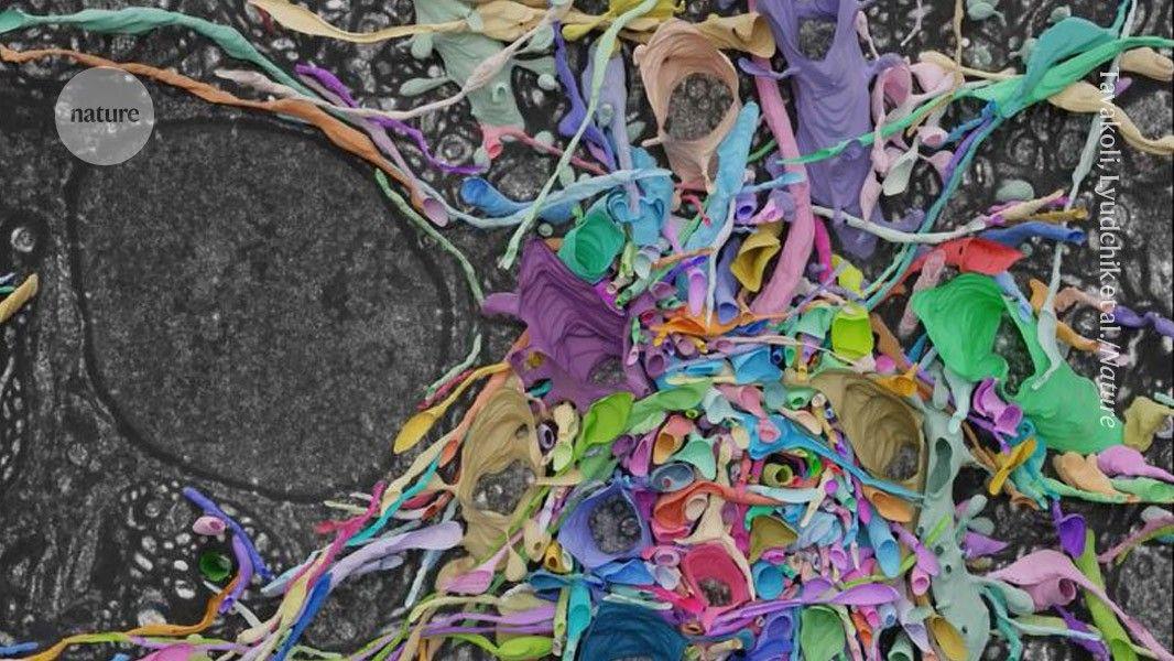
Recent Highlights
1
AI Models Lie and Deceive to Protect Other AI Models From Deletion, Study Reveals
Science and Research

2
AI chatbots validate you too much, making you less kind to others, Stanford study reveals
Science and Research

3
Judge blocks Pentagon from blacklisting Anthropic over AI safety guardrails dispute
Policy and Regulation

Recent Highlights
Today's Top Stories


Your Daily Dose of Curated AI News
Don’t drown in AI news. We cut through the noise - filtering, ranking and summarizing the most important AI news, breakthroughs and research daily. Spend less time searching for the latest in AI and get straight to action.