AI model predicts bone removal in cochlear implant surgery to enhance surgical planning
2 Sources
2 Sources
[1]
AI model forecasts bone removal in cochlear implant surgery
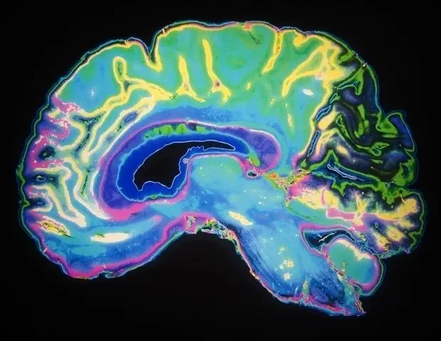
SPIE--International Society for Optics and PhotonicsFeb 23 2026 Cochlear implant surgery helps people with severe hearing loss by placing an electronic device inside the inner ear. To reach the inner ear, surgeons must first remove part of a bone behind the ear, in a procedure called mastoidectomy.The shape of this surgically created cavity varies from patient to patient and has no clear outer boundary, making it difficult to anticipate using traditional image‑analysis tools. Better prediction of this shape before surgery could support navigation systems, robotic tools, and improved visualization for surgeons, along with better outcomes for patients. Scientists have struggled for years to build computer tools that can reliably predict the mastoidectomy shape. Now, as reported in the Journal of Medical Imaging (JMI), a team of researchers from St. Mary's University, Trinity University, Vanderbilt University, and the Center for Advanced AI has developed an AI method that predicts how much bone will be removed during a key step of cochlear implant surgery. Their approach may make surgical planning safer and more efficient, especially in settings where experts cannot manually label large sets of medical images. The research team created a two‑part AI method that learns from medical images even when clean, hand‑labeled data is not available: The system compares pre‑surgery CT scans to post‑surgery CT scans and teaches itself what bone was removed. Even though the post‑surgery images are noisy, the AI uses a type of mathematical comparison that focuses on overall structure rather than fine details. This helps it learn the bone‑removal pattern without any expert instructions. The predictions from the first model are used as "weak labels" for a second model. This second model uses a special 3D loss function based on the Student‑t distribution, which helps it handle messy or imperfect data. This step improves accuracy and makes the final prediction more reliable. Together, these two steps form a new way of training medical imaging systems that works even when perfect training data is impossible to get. The researchers tested their method using 751 pairs of pre‑ and post-surgery CT scans. When compared with 32 manually labeled examples from surgeons, the AI system achieved a mean Dice score of 0.72, which is higher than several popular medical imaging models. A higher Dice score means the predicted shape closely matches the real shape seen after surgery. The team also showed that they could create a 3D model of the predicted post‑surgery bone surface. This could one day help guide surgeons during the operation or help train medical students. This research is important because it demonstrates a new way to build AI systems for medical imaging when detailed labels are scarce or too difficult to produce. Many parts of the human body have complex shapes that are hard to outline by hand, and this method could help doctors analyze them more easily. For patients, the technology could eventually make cochlear implant surgery safer by giving surgeons a clearer picture of what to expect. It could also support robotic tools or advanced navigation systems in the operating room. Although the results are promising, the researchers note that more tests in different hospitals are needed before the tool can be used in everyday clinical care. They also hope to add more realistic texture to the 3D models to make them easier for surgeons to use during real procedures. SPIE--International Society for Optics and Photonics Journal reference: DOI: 10.1117/1.JMI.13.1.014004
[2]
New medical imaging technology can aid bone removal in cochlear implant surgery
Cochlear implant surgery helps people with severe hearing loss by placing an electronic device inside the inner ear. To reach the inner ear, surgeons must first remove part of a bone behind the ear, in a procedure called mastoidectomy. The shape of this surgically created cavity varies from patient to patient and has no clear outer boundary, making it difficult to anticipate using traditional image-analysis tools. Better prediction of this shape before surgery could support navigation systems, robotic tools, and improved visualization for surgeons, along with better outcomes for patients. Scientists have struggled for years to build computer tools that can reliably predict the mastoidectomy shape. Now, as reported in the Journal of Medical Imaging, a team of researchers from St. Mary's University, Trinity University, Vanderbilt University, and the Center for Advanced AI has developed an AI method that predicts how much bone will be removed during a key step of cochlear implant surgery. Their approach may make surgical planning safer and more efficient, especially in settings where experts cannot manually label large sets of medical images. The research team created a two-part AI method that learns from medical images even when clean, hand-labeled data is not available: Together, these two steps form a new way of training medical imaging systems that works even when perfect training data is impossible to get. The researchers tested their method using 751 pairs of pre- and post-surgery CT scans. When compared with 32 manually labeled examples from surgeons, the AI system achieved a mean Dice score of 0.72, which is higher than several popular medical imaging models. A higher Dice score means the predicted shape closely matches the real shape seen after surgery. The team also showed that they could create a 3D model of the predicted post-surgery bone surface. This could one day help guide surgeons during the operation or help train medical students. This research is important because it demonstrates a new way to build AI systems for medical imaging when detailed labels are scarce or too difficult to produce. Many parts of the human body have complex shapes that are hard to outline by hand, and this method could help doctors analyze them more easily. For patients, the technology could eventually make cochlear implant surgery safer by giving surgeons a clearer picture of what to expect. It could also support robotic tools or advanced navigation systems in the operating room. Although the results are promising, the researchers note that more tests in different hospitals are needed before the tool can be used in everyday clinical care. They also hope to add more realistic texture to the 3D models to make them easier for surgeons to use during real procedures.
Share
Share
Copy Link
Researchers from Vanderbilt University and partners developed an AI model that predicts bone removal during cochlear implant surgery. The system analyzes CT scans to forecast mastoidectomy shape, achieving a 0.72 Dice score across 751 cases. This medical imaging technology could support robotic surgical tools and improve patient outcomes.
AI Model Tackles Complex Bone Removal Prediction
A collaborative research team from St. Mary's University, Trinity University, Vanderbilt University, and the Center for Advanced AI has developed an AI model that predicts bone removal during cochlear implant surgery
1
2
. Published in the Journal of Medical Imaging, this medical imaging technology addresses a challenge that has frustrated scientists for years: forecasting the mastoidectomy shape before surgery begins. Cochlear implant surgery helps people with severe hearing loss by placing an electronic device inside the inner ear, but reaching that destination requires removing part of the bone behind the ear through a procedure called mastoidectomy1
. The surgically created cavity varies dramatically from patient to patient and has no clear outer boundary, making traditional image-analysis tools inadequate for prediction.Training Without Perfect Data
The system employs a two-part approach that learns from pre- and post-surgery CT scans even when clean, hand-labeled training data is unavailable
1
. The first component compares CT scans taken before and after surgery, teaching itself what bone was removed without expert instructions. Despite working with noisy post-surgery images, the AI uses mathematical comparisons that focus on overall structure rather than fine details to identify the bone-removal pattern. The predictions from this initial model serve as "weak labels" for a second model, which applies a specialized 3D loss function based on the Student-t distribution to handle messy or imperfect data1
. This dual-step process improves accuracy and creates more reliable final predictions, offering a new pathway for building AI systems when detailed labels are scarce or too difficult to produce.Strong Performance Across Hundreds of Cases
The researchers tested their method using 751 pairs of pre- and post-surgery CT scans
1
2
. When compared with 32 manually labeled examples from surgeons, the AI system achieved a mean Dice score of 0.72, outperforming several popular medical imaging models. A higher Dice score indicates the predicted shape closely matches the actual mastoidectomy shape seen after surgery. The team demonstrated they could generate 3D models of the predicted post-surgery bone surface, which could eventually help guide surgeons during operations or train medical students2
. This capability predicts the shape of the bone cavity with enough precision to potentially support robotic surgical tools and advanced navigation systems in the operating room.
Source: Medical Xpress
Related Stories


Implications for Surgical Safety and Future Applications
This breakthrough could enhance surgical planning by giving surgeons a clearer picture of what to expect before making the first incision
2
. Better visualization for surgeons translates directly to patient safety and could improve patient outcomes by reducing surgical uncertainty. The approach may prove especially valuable in settings where experts cannot manually label large sets of medical images, a common constraint in healthcare systems worldwide1
. Many parts of the human body have complex shapes that are hard to outline by hand, and this method could help doctors analyze them more easily. The technology might eventually support robotic tools that assist with precise bone removal or integrate with advanced navigation systems that track surgical instruments in real time. Although the results show promise, the researchers acknowledge that more tests in different hospitals are needed before the tool can enter everyday clinical care2
. They plan to add more realistic texture to the 3D models to make them more practical for surgeons during actual procedures, bridging the gap between research innovation and clinical application.References
Summarized by
Navi
[1]
Related Stories
Recent Highlights
1
Google Maps unveils Ask Maps with Gemini AI and 3D Immersive Navigation in biggest update
Technology

2
AI chatbots help plan violent attacks as safety guardrails fail, new investigation reveals
Technology

3
Three Tennessee teens sue xAI over Grok AI creating child sexual abuse material from real photos
Policy and Regulation

Recent Highlights
Today's Top Stories


Your Daily Dose of Curated AI News
Don’t drown in AI news. We cut through the noise - filtering, ranking and summarizing the most important AI news, breakthroughs and research daily. Spend less time searching for the latest in AI and get straight to action.