AI Revolutionizes Breast Cancer Detection: New Tools Predict Risk and Catch Missed Tumors
3 Sources
3 Sources
[1]
AI tool identifies women at high risk of interval breast cancer
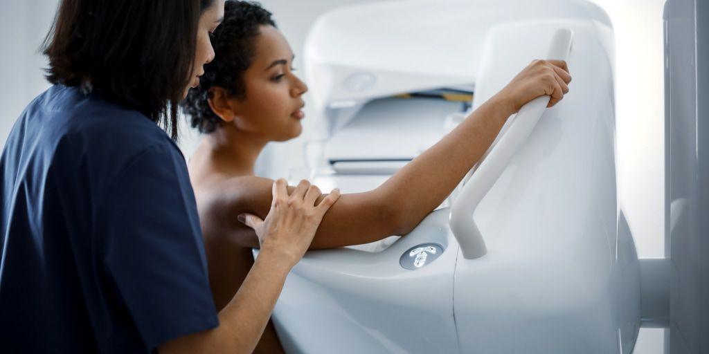
In a study of more than 100,000 screening mammograms, researchers demonstrated the potential of an AI tool to help identify women at higher risk of developing interval breast cancer, which is breast cancer that is diagnosed between regular screening mammograms. Results of the new study were published today in Radiology. "Interval cancers generally have a worse prognosis compared with screen-detected cancers, because they tend to be either larger or more aggressive," said co-author Fiona J. Gilbert, M.B.Ch.B., professor of radiology, Department of Radiology at the University of Cambridge, U.K., and honorary consultant radiologist at Addenbrooke's Hospital. "That's why it's important to minimize the number of interval cancers that you have in any screening program." Using a large retrospective dataset from the U.K.'s triennial screening program, Dr. Gilbert and lead researcher Joshua W. D. Rothwell, an M.B.B.S./Ph.D. student at the University of Cambridge, employed AI to identify women for supplemental imaging to find interval cancers. "Personalized breast cancer screening depends on accurately assessing an individual's risk of developing breast cancer within a specific timeframe," Dr. Gilbert said. "We can use supplemental imaging and adjust screening frequency based on a woman's breast density and likelihood of developing breast cancer within a short timeframe." The study cohort included 134,217 screening mammograms on the same number of women (aged 50-70), with 524 interval cancers. The exams were performed between 2014 and 2016 at two U.K. triennial Breast Screening Program centers using two different mammography systems. Negative (no cancer found) digital screening mammograms were processed by Mirai, a deep learning-based algorithm, producing a generalized risk score for developing an interval breast cancer. The AI tool primarily uses information from the mammogram, including tumor features and breast density, to make a risk prediction. The AI tool's 3-year risk scores retrospectively predicted 3.6% (19/524), 14.5% (76/524), 26.1% (137/524) and 42.4% (222/524) of the 524 interval cancers for women assigned the highest 1%, 5%, 10% and 20% scores. Identification of the interval cancers is equivalent to an additional cancer detection rate of 0.1, 0.6, 1.0, and 1.7 per 1,000. "Our results suggest that further workup of mammograms within the top 20% of scores could yield 42.4% of interval cancers, meaning that Mirai could be used to identify women for supplemental imaging or a shortened screening interval, instead of or in addition to breast density," Rothwell said. The AI tool performed better at predicting interval cancers within a year of the screening examination versus 12 to 24 months or 24 to 36 months later. While the tool was less effective in women with extremely dense breast tissue, it showed superior performance compared to conventional risk prediction tools. In the U.K., 2.2 million women are screened for breast cancer every year. According to Dr. Gilbert, AI could help optimize the country's triennial breast cancer screening program by improving the selection criteria for women who could benefit from supplemental imaging, such as MRI or contrast-enhanced mammography, or by shortening the screening interval. "If we called back 20% of women for supplemental imaging, we'd have to find the capacity to offer contrast-enhanced mammography or MRI to 440,000 women," she said. Next steps for the researchers include comparing commercially available predictive AI tools, conducting economic modeling and a cost-effective analysis, and conducting a trial utilizing predictive AI to identify women most likely to benefit from supplemental breast imaging following screening mammography. "Identifying women at an increased risk of developing breast cancer is a complex, multifactorial problem," Dr. Gilbert said. "The goal is to accurately identify the women most likely to have an interval cancer while minimizing the volume of supplemental imaging performed."
[2]
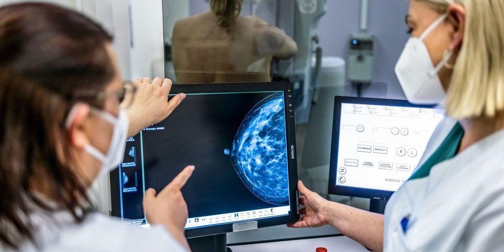
AI mammogram readings are already helping doctors detect breast cancer
Companies help fuel breast cancer research as proposed cuts to federal funding loom When a radiologist reviewed Deirdre Hall's mammogram images last summer, everything seemed fine. There were no shadows or lumps or irregular patches that could signal cancer. The doctor gave it a second look for one reason: artificial intelligence software had drawn a circle around an area in the upper part of her left breast that it found suspicious. Because the AI software had put up that red flag, Hall, 55, got an order for an ultrasound that led to a biopsy. There were four cancerous tumors in the spot AI had identified. "This would have been completely missed without the AI," said Dr. Sean Raj, chief medical officer and chief innovation officer at SimonMed Imaging in Tempe, Arizona, where Hall had her mammogram. Not only was Hall's breast tissue dense, but the layers of tissue crisscrossed over each other in a particularly complicated pattern. "It camouflaged the cancer," said Raj, a breast imaging specialist. "Even I could have missed it." They caught her cancer at Stage 1, said Hall, who's a respiratory therapist at a local hospital. "They didn't find anything in the lymph nodes, which they were grateful for," she said. "I'm so glad they caught it early." When reading women's routine mammograms, radiologists are increasingly augmenting their eyes with artificial intelligence. While many major medical centers have adopted the technology enthusiastically, some experts point to concerns, including a lack of studies in the U.S. showing that AI actually saves lives and does not needlessly raise concerns about benign growths. Experts train AI software by feeding it hundreds of thousands, or sometimes millions of mammogram images. Some of the images contain cancerous tumors, and, over time, the AI learns to distinguish the often subtle differences between malignant and benign tissue. Some AI programs, like the one used on Hall, identify a suspicious area. Others predict the chance that a woman will develop breast cancer. At the University of California, San Francisco, researchers are using AI to try to speed up the time from a mammogram to cancer diagnosis. In a study released this week, the radiologists used the technology to flag suspicious-looking mammograms so those patients could be seen more quickly. For patients with breast cancer, that AI triage cut the average time from mammogram to biopsy by 87%, from 73 days to nine days. The study was posted Tuesday to the preprint server MedRxiv. (Studies posted to preprint servers have not been peer-reviewed.) However, Dr. Sonja Hughes, vice president of community health at Susan G. Komen, a breast cancer organization, said more research is needed before AI is used as the standard of care. "We're not there yet," she said. "We don't have enough research or enough data." Mammograms have saved countless lives, but they're imperfect. Dense breast tissue, which is a risk factor for developing cancer, makes mammograms harder to interpret. About 40% of U.S. women have dense breasts, according to the American Cancer Society. "It's like trying to find a snowball in a blizzard," said Dr. Otis Brawley, a professor of oncology and epidemiology at Johns Hopkins University. The Food and Drug Administration has authorized many AI programs for mammograms, with varying rates of accuracy. The AI software used on Hall's mammogram, called Lunit, accurately identified cancers 88.6% of the time, according to a 2024 JAMA Oncology study of more than 8,800 women in Sweden who got mammograms. Another study published in Radiology noted that AI software caught cancers that were missed by two radiologists. However, in the Sweden study, AI gave a false positive 7% of the time, saying there might be a tumor when there wasn't one. A false positive can trigger more testing and anxiety while waiting for results. With any mammogram, the chance of having a false positive result is about 10%, according to research. Major academic medical centers using AI in their imaging centers include the MD Anderson Cancer Center, the Mount Sinai Hospital in New York City, the Perelman Center for Advanced Medicine at the University of Pennsylvania, the Siteman Cancer Center at Barnes-Jewish Hospital, and MedStar Health. In all centers, the software is used along with, not instead of, a radiologist's eyes, as FDA regulations require a doctor to interpret mammograms. Some breast imaging experts see advantages to this human-machine combination. "The nice thing about AI is that it doesn't get tired," said Dr. Lisa Abramson, associate professor of radiology at Mount Sinai. "It's not going to replace the job or the expertise of radiologists, but I think it's only going to enhance our ability to detect more and more breast cancers." Brawley, the Johns Hopkins professor, said AI could help women who don't have access to radiologists who specialize in breast imaging, and instead have their mammograms read by general radiologists. A study using RadNet's software found that without AI, specialists correctly identified breast cancers 89% of the time, compared with 84% for generalists. With AI, the accuracy for both groups rose to about 93%. "It's incredibly subjective when a human reads a mammogram," Brawley said. "Maybe it's going to reduce the disparities in how these things are read." Typically, academic medical centers don't charge patients extra for the use of AI software, and they can't charge insurance companies for it, since there's no billing code specifically for the AI, according to Susan G. Komen, a nonprofit breast cancer organization. SimonMed, which has centers in 11 states, and RadNet, which has centers in eight states, don't charge for an initial layer of AI on mammograms, although patients are charged $40 and $50 respectively if they opt to have their images run through a second set of the technology. Brawley worries that AI might be too good at its job. According to the American Cancer Society, it's possible that mammograms flag some tumors that are technically cancerous, but not life-threatening. The patient then undergoes the physical, emotional, and financial toll of treating a tumor that was never going to hurt her. "It'scancer, but it's not genetically programmed to grow, spread, or kill," Brawley said. "I am worried that AI may help us find even more of these tumors that don't need to be found." Brawley pointed to the lack of data in the U.S. that shows AI actually saves women's lives. Last month, researchers at the University of California, Los Angeles and University of California, Davis, announced a $16 million, two-year study at seven medical centers to take a deeper look at the technology. There are several other concerns about using AI in mammography. The technology isn't perfect, and some worry that doctors could make mistakes if they become too dependent on it, according to an article last year in RadioGraphics. That's why radiologists emphasize that AI is a tool, not a solution in itself. "It's not going to replace the job or the expertise of radiologists," said Abramson, the breast radiologist at Mount Sinai. "I think it's only going to enhance our ability to detect more and more breast cancer." Another concern is that if AI is trained mainly on breast images of white women, it could be less accurate for women of color, since genetic differences can make tumors look different. Hall, the Arizona patient, said she's not necessarily a fan of AI in general -- she says she finds the technology "creepy" -- but she's glad she paid $50 for the extra AI on her mammogram. "I don't love all this AI stuff, but I definitely love this for me or anyone else in my position," she said. "No matter how it was found, I'm glad it was found." Guidance from the United Services Preventive Services Task Force recommends women to get a mammogram every other year starting at age 40. According to American Cancer Society guidelines: Dr. Shanthi Sivendran, senior vice president at the American Cancer Society, offers guidance for more accurate breast cancer screening.
[3]
EXCLUSIVE: 'Revolutionary' AI-Powered Mammograms Can Predict Breast Cancer Risk
Routine mammograms are the mainstay of breast cancer screening. But, with a new type of analysis powered by artificial intelligence, doctors may be able to also predict whether or not you'll develop breast cancer in the next few years. The technology, called Clairity Breast, is the first of its kind to receive authorization from the Food and Drug Administration. It's just starting to appear in some hospitals in the U.S. And, experts say, when interpreted correctly, it can be an important tool in identifying those with a higher risk for breast cancer. Using images from a mammogram, the technology generates a risk score, which is the probability that someone will develop breast cancer in the next five years. There are other ways that experts can assess that risk, which mainly includes taking note of factors such as a family history of cancer, genetic risks and lifestyle. But recent estimates suggest that the majority of breast cancers are not due to family history or genetic risk factors. Instead of using traditional risk factors to predict risk, Clairity's AI platform was trained on hundreds of thousands of mammograms to look for patterns that would suggest someone is more likely to develop breast cancer. 'The computer can learn the patterns in a mammogram in a woman that will develop breast cancer in the next five years and distinguish those from women that will not," Dr. Connie Lehman, founder of Clairity and chief of breast imaging at Massachusetts General Hospital, told NBC's Savannah Sellers during an Oct. 22, 2025 segment on the TODAY show. And, in a 2022 study in the Journal of the National Cancer Institute, predictions from the Clairity model were often more accurate than traditional risk assessment tools. The development of this new technology marks a "revolutionary" moment, according to Donna McKay, CEO of the Breast Cancer Research Foundation, which funded some of the research that went into Clairity. "This is a paradigm shift from taking what is a mammogram, which is a diagnostic tool, and actually turning it into a predictive tool," McKay said. However, it's important to put the results from Clairity's analysis into the proper context. "This tool is great, and it should be used appropriately, meaning it indicates people who are at high risk," said Dr. Laurie Margolies, vice chair of breast imaging for the Mount Sinai Health Service. "But it doesn't catch everybody who will get cancer in the next five years," Margolies explains. "So we have to be careful that people don't over-rely on it." A Clairity score indicating a high risk doesn't automatically mean someone will develop breast cancer. And a low or average risk doesn't guarantee that they won't. For those with an average Clairity risk score, regular screening (mammograms every one to two years) is appropriate, Lehman said. But for people with a higher risk, MRIs, contrast-enhanced mammograms or ultrasounds would be recommended, she said. And keep in mind that Clairity is still working to get the test covered by insurance. For now, Lehman said, "We really believe that this test for women absolutely can be offered for less than $200." That is in addition to the cost of a mammogram, which is typically covered if it's part of a routine screening. The goal is to reach as many women as possible, especially those who don't think they're at risk, Lehman said. "We're really going to change the face of breast cancer."
Share
Share
Copy Link
Breakthrough AI technologies are transforming breast cancer screening by predicting future cancer risk and identifying tumors that radiologists might miss. Multiple studies show promising results for AI-powered mammogram analysis in improving early detection.
AI Tools Identify High-Risk Women for Interval Breast Cancer
Researchers at the University of Cambridge have demonstrated the potential of artificial intelligence to identify women at higher risk of developing interval breast cancer—cancers that emerge between regular screening mammograms. In a comprehensive study of 134,217 screening mammograms from women aged 50-70, the AI tool Mirai successfully predicted interval cancers with remarkable precision
1
.
Source: NBC
The study, published in Radiology, revealed that AI could identify 42.4% of interval cancers by focusing on the top 20% of risk scores. "Interval cancers generally have a worse prognosis compared with screen-detected cancers, because they tend to be either larger or more aggressive," explained co-author Professor Fiona J. Gilbert from the University of Cambridge
1
.Real-World Success Stories in Cancer Detection
The practical impact of AI-assisted mammogram reading is already saving lives in clinical settings. Deirdre Hall, a 55-year-old respiratory therapist, experienced this firsthand when AI software flagged a suspicious area in her mammogram that radiologists had initially deemed normal. The AI's intervention led to the discovery of four Stage 1 cancerous tumors that would have otherwise been missed
2
.
Source: TODAY.com
"This would have been completely missed without the AI," said Dr. Sean Raj, chief medical officer at SimonMed Imaging, where Hall received her screening. The case highlights how AI can detect cancers camouflaged by dense breast tissue, which affects approximately 40% of U.S. women and makes mammogram interpretation particularly challenging
2
.FDA-Approved Predictive Technology Enters Market
Clairity Breast has become the first AI technology of its kind to receive FDA authorization for predicting breast cancer risk. Unlike traditional risk assessment methods that rely on family history and genetic factors, Clairity analyzes mammogram images to generate a probability score for developing breast cancer within the next five years
3
.Dr. Connie Lehman, founder of Clairity and chief of breast imaging at Massachusetts General Hospital, explained that "the computer can learn the patterns in a mammogram in a woman that will develop breast cancer in the next five years and distinguish those from women that will not" .
Related Stories


Performance Metrics and Clinical Implementation
Studies demonstrate varying but promising accuracy rates for AI mammogram analysis. The Lunit software used in Hall's case achieved 88.6% accuracy in identifying cancers, according to a 2024 JAMA Oncology study of over 8,800 women in Sweden. However, the technology also produced false positives 7% of the time, compared to the typical 10% false positive rate for standard mammograms
2
.At UC San Francisco, researchers have used AI to dramatically reduce diagnosis timelines, cutting the average time from mammogram to biopsy by 87%—from 73 days to just nine days for cancer patients
2
.Widespread Adoption Across Medical Centers
Major academic medical centers are increasingly integrating AI into their breast imaging programs. Institutions including MD Anderson Cancer Center, Mount Sinai Hospital, University of Pennsylvania's Perelman Center, and MedStar Health have adopted AI-assisted mammogram reading
2
.
Source: Medical Xpress
"The nice thing about AI is that it doesn't get tired," noted Dr. Lisa Abramson, associate professor of radiology at Mount Sinai. "It's not going to replace the job or the expertise of radiologists, but I think it's only going to enhance our ability to detect more and more breast cancers"
2
.References
Summarized by
Navi
[1]
Related Stories
Recent Highlights


Recent Highlights
Today's Top Stories


Your Daily Dose of Curated AI News
Don’t drown in AI news. We cut through the noise - filtering, ranking and summarizing the most important AI news, breakthroughs and research daily. Spend less time searching for the latest in AI and get straight to action.