AI Tool Predicts Pediatric Brain Cancer Recurrence with High Accuracy
3 Sources
3 Sources
[1]
Artificial intelligence tool helps predict relapse of pediatric brain cancer
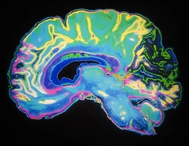
Artificial intelligence (AI) shows tremendous promise for analyzing vast medical imaging datasets and identifying patterns that may be missed by human observers. AI-assisted interpretation of brain scans may help improve care for children with brain tumors called gliomas, which are typically treatable but vary in risk of recurrence. Investigators from Mass General Brigham and collaborators at Boston Children's Hospital and Dana-Farber/Boston Children's Cancer and Blood Disorders Center trained deep learning algorithms to analyze sequential, post-treatment brain scans and flag patients at risk of cancer recurrence. Their results are published in The New England Journal of Medicine AI. "Many pediatric gliomas are curable with surgery alone, but when relapses occur, they can be devastating," said corresponding author Benjamin Kann, MD, of the Artificial Intelligence in Medicine (AIM) Program at Mass General Brigham and the Department of Radiation Oncology at Brigham and Women's Hospital. "It is very difficult to predict who may be at risk of recurrence, so patients undergo frequent follow-up with magnetic resonance (MR) imaging for many years, a process that can be stressful and burdensome for children and families. We need better tools to identify early which patients are at the highest risk of recurrence." Studies of relatively rare diseases, like pediatric cancers, can be challenged by limited data. This study, which was funded in part by the National Institutes of Health, leveraged institutional partnerships across the country to collect nearly 4,000 MR scans from 715 pediatric patients. To maximize what AI could "learn" from a patient's brain scans -- and more accurately predict recurrence -- the researchers employed a technique called temporal learning, which trains the model to synthesize findings from multiple brain scans taken over the course of several months post-surgery. Typically, AI models for medical imaging are trained to draw conclusions from single scans; with temporal learning, which has not previously been used for medical imaging AI research, images acquired over time inform the algorithm's prediction of cancer recurrence. To develop the temporal learning model, the researchers first trained the model to sequence a patient's post-surgery MR scans in chronological order so that the model could learn to recognize subtle changes. From there, the researchers fine-tuned the model to correctly associate changes with subsequent cancer recurrence, where appropriate. Ultimately, the researchers found that the temporal learning model predicted recurrence of either low- or high-grade glioma by one-year post-treatment, with an accuracy of 75-89 percent -- substantially better than the accuracy associated with predictions based on single images, which they found to be roughly 50 percent (no better than chance). Providing the AI with images from more timepoints post-treatment increased the model's prediction accuracy, but only four to six images were required before this improvement plateaued. The researchers caution that further validation across additional settings is necessary prior to clinical application. Ultimately, they hope to launch clinical trials to see if AI-informed risk predictions can result in improvements to care -- whether by reducing imaging frequency for the lowest-risk patients or by preemptively treating high-risk patients with targeted adjuvant therapies. "We have shown that AI is capable of effectively analyzing and making predictions from multiple images, not just single scans," said first author Divyanshu Tak, MS, of the AIM Program at Mass General Brigham and the Department of Radiation Oncology at the Brigham. "This technique may be applied in many settings where patients get serial, longitudinal imaging, and we're excited to see what this project will inspire." Authorship: In addition to Kann and Tak, Mass General Brigham authors include Biniam A. Garomsa, Anna Zapaishchykova, Zezhong Ye, Maryam Mahootiha, Tafadzwa Chaunzwa, Hugo JWL Aerts, and Daphne Haas-Kogan. Additional authors include Sridhar Vajapeyam, Juan Carlos Climent Pardo, Ceilidh Smith, Ariana M. Familiar, Kevin X. Liu, Sanjay Prabhu, Pratiti Bandopadhayay, Ali Nabavizadeh, Sabine Mueller, and Tina Y. Poussaint, Funding: This study was supported in part by the National Institute of Health/ the National Cancer Institute (NIH/NCI) (U54 CA274516 and P50 CA165962), and Botha-Chan Low Grade Glioma Consortium. We would also like to acknowledge the Children's Brain Tumor Network (CBTN) for the imaging and clinical data access.
[2]
AI predicts pediatric glioma recurrence using multiple brain scans
Mass General BrighamApr 24 2025 Artificial intelligence (AI) shows tremendous promise for analyzing vast medical imaging datasets and identifying patterns that may be missed by human observers. AI-assisted interpretation of brain scans may help improve care for children with brain tumors called gliomas, which are typically treatable but vary in risk of recurrence. Investigators from Mass General Brigham and collaborators at Boston Children's Hospital and Dana-Farber/Boston Children's Cancer and Blood Disorders Center trained deep learning algorithms to analyze sequential, post-treatment brain scans and flag patients at risk of cancer recurrence. Their results are published in The New England Journal of Medicine AI. Many pediatric gliomas are curable with surgery alone, but when relapses occur, they can be devastating. It is very difficult to predict who may be at risk of recurrence, so patients undergo frequent follow-up with magnetic resonance (MR) imaging for many years, a process that can be stressful and burdensome for children and families. We need better tools to identify early which patients are at the highest risk of recurrence." Benjamin Kann, MD, corresponding author of the Artificial Intelligence in Medicine (AIM) Program at Mass General Brigham and the Department of Radiation Oncology at Brigham and Women's Hospital Studies of relatively rare diseases, like pediatric cancers, can be challenged by limited data. This study, which was funded in part by the National Institutes of Health, leveraged institutional partnerships across the country to collect nearly 4,000 MR scans from 715 pediatric patients. To maximize what AI could "learn" from a patient's brain scans - and more accurately predict recurrence - the researchers employed a technique called temporal learning, which trains the model to synthesize findings from multiple brain scans taken over the course of several months post-surgery. Typically, AI models for medical imaging are trained to draw conclusions from single scans; with temporal learning, which has not previously been used for medical imaging AI research, images acquired over time inform the algorithm's prediction of cancer recurrence. To develop the temporal learning model, the researchers first trained the model to sequence a patient's post-surgery MR scans in chronological order so that the model could learn to recognize subtle changes. From there, the researchers fine-tuned the model to correctly associate changes with subsequent cancer recurrence, where appropriate. Ultimately, the researchers found that the temporal learning model predicted recurrence of either low- or high-grade glioma by one-year post-treatment, with an accuracy of 75-89 percent - substantially better than the accuracy associated with predictions based on single images, which they found to be roughly 50 percent (no better than chance). Providing the AI with images from more timepoints post-treatment increased the model's prediction accuracy, but only four to six images were required before this improvement plateaued. The researchers caution that further validation across additional settings is necessary prior to clinical application. Ultimately, they hope to launch clinical trials to see if AI-informed risk predictions can result in improvements to care - whether by reducing imaging frequency for the lowest-risk patients or by preemptively treating high-risk patients with targeted adjuvant therapies. "We have shown that AI is capable of effectively analyzing and making predictions from multiple images, not just single scans," said first author Divyanshu Tak, MS, of the AIM Program at Mass General Brigham and the Department of Radiation Oncology at the Brigham. "This technique may be applied in many settings where patients get serial, longitudinal imaging, and we're excited to see what this project will inspire." Mass General Brigham Journal reference: Tak, D., et al. (2025) Longitudinal Risk Prediction for Pediatric Glioma with Temporal Deep Learning. NEJM AI. doi.org/10.1056/AIoa2400703.
[3]
AI Predicts Brain Cancer Recurrence in Kids - Neuroscience News
Summary: Researchers have developed an AI model that analyzes sequences of brain scans to accurately predict tumor recurrence in children with gliomas. By applying a method called temporal learning, the model interprets subtle changes in MR images taken post-treatment over time. The study found that using multiple images significantly outperforms traditional single-scan analysis, reaching prediction accuracy rates as high as 89%. This approach could one day reduce the burden of frequent scans or enable earlier interventions. Artificial intelligence (AI) shows tremendous promise for analyzing vast medical imaging datasets and identifying patterns that may be missed by human observers. AI-assisted interpretation of brain scans may help improve care for children with brain tumors called gliomas, which are typically treatable but vary in risk of recurrence. Investigators from Mass General Brigham and collaborators at Boston Children's Hospital and Dana-Farber/Boston Children's Cancer and Blood Disorders Center trained deep learning algorithms to analyze sequential, post-treatment brain scans and flag patients at risk of cancer recurrence. Their results are published in The New England Journal of Medicine AI. "Many pediatric gliomas are curable with surgery alone, but when relapses occur, they can be devastating," said corresponding author Benjamin Kann, MD, of the Artificial Intelligence in Medicine (AIM) Program at Mass General Brigham and the Department of Radiation Oncology at Brigham and Women's Hospital. "It is very difficult to predict who may be at risk of recurrence, so patients undergo frequent follow-up with magnetic resonance (MR) imaging for many years, a process that can be stressful and burdensome for children and families. We need better tools to identify early which patients are at the highest risk of recurrence." Studies of relatively rare diseases, like pediatric cancers, can be challenged by limited data. This study, which was funded in part by the National Institutes of Health, leveraged institutional partnerships across the country to collect nearly 4,000 MR scans from 715 pediatric patients. To maximize what AI could "learn" from a patient's brain scans -- and more accurately predict recurrence -- the researchers employed a technique called temporal learning, which trains the model to synthesize findings from multiple brain scans taken over the course of several months post-surgery. Typically, AI models for medical imaging are trained to draw conclusions from single scans; with temporal learning, which has not previously been used for medical imaging AI research, images acquired over time inform the algorithm's prediction of cancer recurrence. To develop the temporal learning model, the researchers first trained the model to sequence a patient's post-surgery MR scans in chronological order so that the model could learn to recognize subtle changes. From there, the researchers fine-tuned the model to correctly associate changes with subsequent cancer recurrence, where appropriate. Ultimately, the researchers found that the temporal learning model predicted recurrence of either low- or high-grade glioma by one-year post-treatment, with an accuracy of 75-89 percent -- substantially better than the accuracy associated with predictions based on single images, which they found to be roughly 50 percent (no better than chance). Providing the AI with images from more timepoints post-treatment increased the model's prediction accuracy, but only four to six images were required before this improvement plateaued. The researchers caution that further validation across additional settings is necessary prior to clinical application. Ultimately, they hope to launch clinical trials to see if AI-informed risk predictions can result in improvements to care -- whether by reducing imaging frequency for the lowest-risk patients or by preemptively treating high-risk patients with targeted adjuvant therapies. "We have shown that AI is capable of effectively analyzing and making predictions from multiple images, not just single scans," said first author Divyanshu Tak, MS, of the AIM Program at Mass General Brigham and the Department of Radiation Oncology at the Brigham. "This technique may be applied in many settings where patients get serial, longitudinal imaging, and we're excited to see what this project will inspire." Authorship: In addition to Kann and Tak, Mass General Brigham authors include Biniam A. Garomsa, Anna Zapaishchykova, Zezhong Ye, Maryam Mahootiha, Tafadzwa Chaunzwa, Hugo JWL Aerts, and Daphne Haas-Kogan. Additional authors include Sridhar Vajapeyam, Juan Carlos Climent Pardo, Ceilidh Smith, Ariana M. Familiar, Kevin X. Liu, Sanjay Prabhu, Pratiti Bandopadhayay, Ali Nabavizadeh, Sabine Mueller, and Tina Y. Poussaint, Funding: This study was supported in part by the National Institute of Health/ the National Cancer Institute (NIH/NCI) (U54 CA274516 and P50 CA165962), and Botha-Chan Low Grade Glioma Consortium. We would also like to acknowledge the Children's Brain Tumor Network (CBTN) for the imaging and clinical data access. Longitudinal Risk Prediction for Pediatric Glioma with Temporal Deep Learning Pediatric glioma recurrence can cause morbidity and mortality; however, recurrence patterns and severity are heterogeneous and challenging to predict with established clinical and genomic markers. As a result, almost all children undergo frequent, long-term, magnetic resonance imaging (MRI) brain surveillance regardless of individual recurrence risk. Longitudinal deep-learning analysis of serial MRI scans may be an effective approach for improving individualized recurrence prediction in gliomas and other cancers, but, thus far, progress has been limited by data availability and current machine-learning approaches. We developed a self-supervised temporal deep-learning approach tailored for longitudinal medical imaging analysis, wherein a multistep model encodes patients' serial MRI scans and is trained to classify the correct chronological order as a pretext task. The pretrained model is then fine-tuned to predict the primary end point of interest -- in this case, 1-year recurrence prediction for pediatric gliomas from the point of last scan -- by leveraging a patient's historical postoperative surveillance scans. We apply the model across 3994 scans from 715 patients followed at three separate institutions in the setting of pediatric low- and high-grade gliomas. Longitudinal imaging analysis with temporal learning improved recurrence prediction performance (F1 score) by up to 58.5% (range, 6.6 to 58.5%) compared with traditional approaches across datasets, with performance improvements in both low- and high-grade gliomas and area under the receiver operating characteristic curve of (range, 75 to 89%) across all datasets. Recurrence prediction performance increased incrementally with the number of historical scans available per patient, reaching plateaus between three and six scans, depending on the dataset. Temporal deep learning enables high-performing longitudinal medical imaging analysis and point-of-care decision support for pediatric brain tumors. Temporal learning may be broadly adaptable to track and predict risk in patients with other cancers and chronic diseases undergoing surveillance imaging. (Funded in part by the National Institutes of Health/National Cancer Institute (U54 CA274516 and P50 CA165962), and Botha-Chan Low Grade Glioma Consortium.)
Share
Share
Copy Link
Researchers develop an AI model using temporal learning to analyze sequential brain scans, predicting glioma recurrence in children with up to 89% accuracy. This breakthrough could revolutionize patient care and treatment strategies.

AI Breakthrough in Predicting Pediatric Brain Cancer Recurrence
Researchers from Mass General Brigham, in collaboration with Boston Children's Hospital and Dana-Farber/Boston Children's Cancer and Blood Disorders Center, have developed a groundbreaking artificial intelligence (AI) tool that significantly improves the prediction of brain cancer recurrence in children. The study, published in The New England Journal of Medicine AI, demonstrates the potential of AI in revolutionizing pediatric cancer care
1
.The Challenge of Pediatric Gliomas
Pediatric gliomas, while often curable with surgery, pose a significant challenge due to the unpredictable nature of their recurrence. Dr. Benjamin Kann, the study's corresponding author, explains, "Many pediatric gliomas are curable with surgery alone, but when relapses occur, they can be devastating"
2
. The current practice involves frequent follow-up magnetic resonance (MR) imaging for years, which can be stressful and burdensome for children and their families.Innovative Temporal Learning Approach
The researchers employed a novel technique called temporal learning to train deep learning algorithms. This method analyzes sequential, post-treatment brain scans to identify subtle changes over time. Unlike traditional AI models that focus on single scans, this approach synthesizes findings from multiple brain scans taken over several months post-surgery
3
.Impressive Accuracy in Predicting Recurrence
The temporal learning model demonstrated remarkable accuracy in predicting glioma recurrence:
- Prediction accuracy of 75-89% for both low- and high-grade gliomas by one-year post-treatment.
- Significantly outperformed single-image based predictions, which had an accuracy of about 50% (no better than chance).
- Optimal performance achieved with 4-6 images, after which improvement plateaued
1
.
Data Collection and Model Development
The study leveraged institutional partnerships across the country to collect nearly 4,000 MR scans from 715 pediatric patients. This extensive dataset was crucial in training the AI model effectively. The researchers first trained the model to sequence post-surgery MR scans chronologically, enabling it to recognize subtle changes. Subsequently, they fine-tuned the model to associate these changes with cancer recurrence
2
.Related Stories

Potential Impact on Patient Care
While further validation is necessary before clinical application, the researchers are optimistic about the potential impact of this AI tool:
- Possibility of reducing imaging frequency for low-risk patients.
- Enabling preemptive treatment with targeted adjuvant therapies for high-risk patients.
- Alleviating the stress and burden of frequent scans on children and families
3
.
Future Directions
The success of this AI model opens up new possibilities in medical imaging analysis. Divyanshu Tak, the study's first author, notes, "This technique may be applied in many settings where patients get serial, longitudinal imaging, and we're excited to see what this project will inspire"
1
. The researchers aim to launch clinical trials to evaluate how AI-informed risk predictions can improve patient care and outcomes.References
Summarized by
Navi
[3]
Related Stories
AI models achieve breakthrough accuracy in diagnosing brain tumors from MRI scans
08 Dec 2025•Health
AI learns to identify childhood cancer survivors who need extra support through patient conversations
28 Mar 2026•Health

AI Model FastGlioma Revolutionizes Brain Tumor Detection During Surgery
14 Nov 2024•Health

Recent Highlights
1
AI chatbots validate you too much, making you less kind to others, Stanford study reveals
Science and Research

2
Anthropic's Claude Code Source Leak Reveals Hidden AI Agent Plans and Extensive System Access
Technology

3
Judge blocks Pentagon from branding Anthropic a security risk over AI safety guardrails dispute
Policy and Regulation

Recent Highlights
Today's Top Stories


Your Daily Dose of Curated AI News
Don’t drown in AI news. We cut through the noise - filtering, ranking and summarizing the most important AI news, breakthroughs and research daily. Spend less time searching for the latest in AI and get straight to action.