LILAC: A Versatile AI System for Analyzing Medical Image Series
3 Sources
3 Sources
[1]
A versatile AI system for analyzing series of medical images
A new AI-based system for analyzing images taken over time can accurately detect changes and predict outcomes, according to a study led by investigators at Weill Cornell Medicine, Cornell's Ithaca campus and Cornell Tech. The system's sensitivity and flexibility could make it useful across a wide range of medical and scientific applications. The new system, termed LILAC (Learning-based Inference of Longitudinal imAge Changes), is based on an AI approach called machine learning. In the study, which appears Feb. 20 in the Proceedings of the National Academy of Sciences, the researchers developed the system and demonstrated it on diverse time-series of images -- also called "longitudinal" image series -- covering developing IVF embryos, healing tissue after wounds and aging brains. The researchers showed that LILAC has a broad ability to identify even very subtle differences between images taken at different times, and to predict related outcome measures such as cognitive scores from brain scans. "This new tool will allow us to detect and quantify clinically relevant changes over time in ways that weren't possible before, and its flexibility means that it can be applied off-the-shelf to virtually any longitudinal imaging dataset," said study senior author Dr. Mert Sabuncu, vice chair of research and a professor of electrical engineering in radiology at Weill Cornell Medicine and professor in the School of Electrical and Computer Engineering at Cornell University's Ithaca campus and Cornell Tech. The study's first author is Dr. Heejong Kim, an instructor of artificial intelligence in radiology at Weill Cornell Medicine and a member of the Sabuncu Laboratory. Traditional methods for analyzing longitudinal image datasets tend to require extensive customization and pre-processing. For example, researchers studying the brain may take raw brain MRI data and pre-process the image data to focus on just one brain area, also correcting for different view angles, sizing differences and other artifacts in the data -- all before performing the main analysis. The researchers designed LILAC to work much more flexibly, in effect automatically performing such corrections and finding relevant changes. "This enables LILAC to be useful not just across different imaging contexts but also in situations where you aren't sure what kind of change to expect," said Dr. Kim, LILAC's principal designer. In one proof-of-concept demonstration, the researchers trained LILAC on hundreds of sequences of microscope images showing in-vitro-fertilized embryos as they develop, and then tested it against new embryo image sequences. LILAC had to determine, for randomized pairs of images from a given sequence, which image was taken earlier -- a task that cannot be done reliably unless the image data contain a true "signal" indicating time-related change. LILAC performed this task with about 99% accuracy, the few errors occurring in image pairs with relatively short time intervals. LILAC also was highly accurate in ordering pairs of images of healing tissue from the same sequences, and in detecting group-level differences in healing rates between untreated tissue and tissue that received an experimental treatment. Similarly, LILAC predicted the time intervals between MRI images of healthy older adults' brains, as well as individual cognitive scores from MRIs of patients with mild cognitive impairment -- in both cases with much less error compared with baseline methods. The researchers showed in all these cases that LILAC can be adapted easily to highlight the image features that are most relevant for detecting changes in individuals or differences between groups -- which could provide new clinical and even scientific insights. "We expect this tool to be useful especially in cases where we lack knowledge about the process being studied, and where there is a lot of variability across individuals," Dr. Sabuncu said. The researchers now plan to demonstrate LILAC in a real-world setting to predict treatment responses from MRI scans of prostate cancer patients.
[2]
Versatile AI system can detect subtle changes in series of medical images
A new AI-based system for analyzing images taken over time can accurately detect changes and predict outcomes, according to a study led by investigators at Weill Cornell Medicine, Cornell's Ithaca campus and Cornell Tech. The system's sensitivity and flexibility could make it useful across a wide range of medical and scientific applications. The new system, termed LILAC (Learning-based Inference of Longitudinal imAge Changes), is based on an AI approach called machine learning. In the study, which appeared Feb. 20 in the Proceedings of the National Academy of Sciences, the researchers developed the system and demonstrated it on diverse time-series of images -- also called "longitudinal" image series -- covering developing IVF embryos, healing tissue after wounds and aging brains. The researchers showed that LILAC has a broad ability to identify even very subtle differences between images taken at different times, and to predict related outcome measures such as cognitive scores from brain scans. "This new tool will allow us to detect and quantify clinically relevant changes over time in ways that weren't possible before, and its flexibility means that it can be applied off-the-shelf to virtually any longitudinal imaging dataset," said study senior author Dr. Mert Sabuncu, vice chair of research and a professor of electrical engineering in radiology at Weill Cornell Medicine and professor in the School of Electrical and Computer Engineering at Cornell University's Ithaca campus and Cornell Tech. The study's first author is Dr. Heejong Kim, an instructor of artificial intelligence in radiology at Weill Cornell Medicine and a member of the Sabuncu Laboratory. Traditional methods for analyzing longitudinal image datasets tend to require extensive customization and pre-processing. For example, researchers studying the brain may take raw brain MRI data and pre-process the image data to focus on just one brain area, also correcting for different view angles, sizing differences and other artifacts in the data -- all before performing the main analysis. The researchers designed LILAC to work much more flexibly, in effect automatically performing such corrections and finding relevant changes. "This enables LILAC to be useful not just across different imaging contexts but also in situations where you aren't sure what kind of change to expect," said Dr. Kim, LILAC's principal designer. In one proof-of-concept demonstration, the researchers trained LILAC on hundreds of sequences of microscope images showing in-vitro-fertilized embryos as they develop, and then tested it against new embryo image sequences. LILAC had to determine, for randomized pairs of images from a given sequence, which image was taken earlier -- a task that cannot be done reliably unless the image data contain a true "signal" indicating time-related change. LILAC performed this task with about 99% accuracy, the few errors occurring in image pairs with relatively short time intervals. LILAC was also highly accurate in ordering pairs of images of healing tissue from the same sequences, and in detecting group-level differences in healing rates between untreated tissue and tissue that received an experimental treatment. Similarly, LILAC predicted the time intervals between MRI images of healthy older adults' brains, as well as individual cognitive scores from MRIs of patients with mild cognitive impairment -- in both cases with much less error compared with baseline methods. The researchers showed in all these cases that LILAC can be adapted easily to highlight the image features that are most relevant for detecting changes in individuals or differences between groups -- which could provide new clinical and even scientific insights. "We expect this tool to be useful especially in cases where we lack knowledge about the process being studied, and where there is a lot of variability across individuals," Dr. Sabuncu said. The researchers now plan to demonstrate LILAC in a real-world setting to predict treatment responses from MRI scans of prostate cancer patients.
[3]
AI system accurately detects changes in longitudinal medical images
Weill Cornell MedicineFeb 27 2025 A new AI-based system for analyzing images taken over time can accurately detect changes and predict outcomes, according to a study led by investigators at Weill Cornell Medicine, Cornell's Ithaca campus and Cornell Tech. The system's sensitivity and flexibility could make it useful across a wide range of medical and scientific applications. The new system, termed LILAC (Learning-based Inference of Longitudinal imAge Changes), is based on an AI approach called machine learning. In the study, which appears Feb. 20 in the Proceedings of the National Academy of Sciences, the researchers developed the system and demonstrated it on diverse time-series of images-also called "longitudinal" image series-covering developing IVF embryos, healing tissue after wounds and aging brains. The researchers showed that LILAC has a broad ability to identify even very subtle differences between images taken at different times, and to predict related outcome measures such as cognitive scores from brain scans. "This new tool will allow us to detect and quantify clinically relevant changes over time in ways that weren't possible before, and its flexibility means that it can be applied off-the-shelf to virtually any longitudinal imaging dataset," said study senior author Dr. Mert Sabuncu, vice chair of research and a professor of electrical engineering in radiology at Weill Cornell Medicine and professor in the School of Electrical and Computer Engineering at Cornell University's Ithaca campus and Cornell Tech. The study's first author is Dr. Heejong Kim, an instructor of artificial intelligence in radiology at Weill Cornell Medicine and a member of the Sabuncu Laboratory. Traditional methods for analyzing longitudinal image datasets tend to require extensive customization and pre-processing. For example, researchers studying the brain may take raw brain MRI data and pre-process the image data to focus on just one brain area, also correcting for different view angles, sizing differences and other artifacts in the data-all before performing the main analysis. The researchers designed LILAC to work much more flexibly, in effect automatically performing such corrections and finding relevant changes. This enables LILAC to be useful not just across different imaging contexts but also in situations where you aren't sure what kind of change to expect. Dr. Heejong Kim, LILAC's principal designer, instructor of artificial intelligence in radiology at Weill Cornell Medicine In one proof-of-concept demonstration, the researchers trained LILAC on hundreds of sequences of microscope images showing in-vitro-fertilized embryos as they develop, and then tested it against new embryo image sequences. LILAC had to determine, for randomized pairs of images from a given sequence, which image was taken earlier-a task that cannot be done reliably unless the image data contain a true "signal" indicating time-related change. LILAC performed this task with about 99% accuracy, the few errors occurring in image pairs with relatively short time intervals. LILAC also was highly accurate in ordering pairs of images of healing tissue from the same sequences, and in detecting group-level differences in healing rates between untreated tissue and tissue that received an experimental treatment. Similarly, LILAC predicted the time intervals between MRI images of healthy older adults' brains, as well as individual cognitive scores from MRIs of patients with mild cognitive impairment-in both cases with much less error compared with baseline methods. The researchers showed in all these cases that LILAC can be adapted easily to highlight the image features that are most relevant for detecting changes in individuals or differences between groups-which could provide new clinical and even scientific insights. "We expect this tool to be useful especially in cases where we lack knowledge about the process being studied, and where there is a lot of variability across individuals," Dr. Sabuncu said. The researchers now plan to demonstrate LILAC in a real-world setting to predict treatment responses from MRI scans of prostate cancer patients. Weill Cornell Medicine Journal reference: Kim, H., et al. (2025). Learning-based inference of longitudinal image changes: Applications in embryo development, wound healing, and aging brain. Proceedings of the National Academy of Sciences. doi.org/10.1073/pnas.2411492122.
Share
Share
Copy Link
Researchers at Weill Cornell Medicine, Cornell's Ithaca campus, and Cornell Tech have developed LILAC, an AI-based system that can accurately detect changes and predict outcomes from longitudinal medical image series.

LILAC: A Breakthrough in Medical Image Analysis
Researchers at Weill Cornell Medicine, Cornell's Ithaca campus, and Cornell Tech have developed a groundbreaking AI system called LILAC (Learning-based Inference of Longitudinal imAge Changes) that promises to revolutionize the analysis of medical image series. This versatile tool, based on machine learning, has demonstrated remarkable accuracy in detecting changes and predicting outcomes across various medical applications
1
.Innovative Approach to Image Analysis
LILAC stands out from traditional methods by offering unprecedented flexibility and sensitivity. Unlike conventional approaches that require extensive customization and pre-processing, LILAC can automatically perform necessary corrections and identify relevant changes
2
. This capability makes it applicable to a wide range of medical and scientific scenarios, particularly where the expected changes are uncertain or highly variable across individuals.Impressive Performance Across Multiple Applications
The research team, led by Dr. Mert Sabuncu and Dr. Heejong Kim, demonstrated LILAC's effectiveness through several proof-of-concept tests:
-
IVF Embryo Development: LILAC achieved 99% accuracy in determining the chronological order of embryo images
3
. -
Wound Healing: The system accurately ordered images of healing tissue and detected differences in healing rates between treated and untreated tissues
1
. -
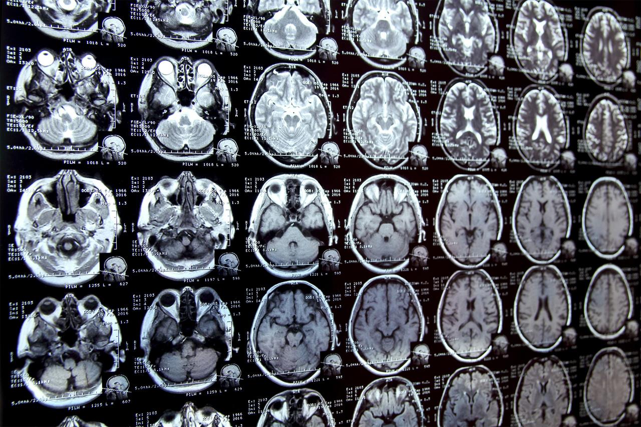
Brain Aging: LILAC predicted time intervals between MRI images of healthy older adults' brains and cognitive scores from MRIs of patients with mild cognitive impairment, outperforming baseline methods
2
.
Potential for Clinical and Scientific Insights
One of LILAC's key strengths is its ability to highlight image features most relevant for detecting changes in individuals or differences between groups. This capability could provide valuable insights for both clinical practice and scientific research
3
.Related Stories

Future Applications
The research team is now planning to demonstrate LILAC's capabilities in a real-world setting, focusing on predicting treatment responses from MRI scans of prostate cancer patients
1
. This application could potentially improve treatment planning and patient outcomes in oncology.Implications for Medical Research and Practice
LILAC's development represents a significant advancement in medical image analysis. Its ability to work across different imaging contexts and detect subtle changes over time could accelerate research in various fields of medicine and biology. Moreover, its flexibility and ease of use make it a promising tool for clinical applications, potentially improving diagnostic accuracy and treatment monitoring
2
.References
Summarized by
Navi
[1]
Related Stories
BiomedParse: A Breakthrough AI Model for Analyzing Multiple Medical Image Types
19 Nov 2024•Health

AI Breakthrough: New Tool Revolutionizes Detection of Rare Gastrointestinal Diseases
25 Oct 2024•Health

MIT's AI System Revolutionizes Medical Image Segmentation for Clinical Research
25 Sept 2025•Science and Research
Recent Highlights
1
Google releases Gemma 4 with Apache 2.0 license, enabling unrestricted local AI on devices
Technology

2
AI Models Lie and Deceive to Protect Other AI Models From Deletion, Study Reveals
Science and Research

3
OpenAI closes $122 billion funding round amid fierce AI competition and profitability questions
Startups

Recent Highlights
Today's Top Stories


Your Daily Dose of Curated AI News
Don’t drown in AI news. We cut through the noise - filtering, ranking and summarizing the most important AI news, breakthroughs and research daily. Spend less time searching for the latest in AI and get straight to action.