AI-Powered Smart Bandage Revolutionizes Wound Healing with 25% Faster Results
4 Sources
4 Sources
[1]
Towards adaptive bioelectronic wound therapy with integrated real-time diagnostics and machine learning-driven closed-loop control - npj Biomedical Innovations
The a-Heal platform includes two main components: the a-Heal wearable device (Fig. 1 and S1), which monitors wounds and delivers on-demand therapy, and the a-Heal ML Physician, a ML driven adaptive diagnostic and treatment algorithm with a graphical user interface (GUI) for human physician oversight, if needed (Fig. 1a). The closed-loop adaptive diagnostics and therapeutic process begins when the onboard camera of the a-Heal wearable device captures an image of the wound and transmits it wirelessly to the a-Heal ML Physician. The ML Physician analyzes the image, generates a diagnosis of wound stage, and prescribes a treatment plan to accelerate wound healing. The wearable device receives the prescribed therapy wirelessly and implements it using bioelectronic actuators that deliver an electric field (EF) or a drug of choice (Fig. 1a). After the therapy takes effect for a specified duration, the wearable device captures a new wound image and restarts the diagnostic and therapeutic cycle (Fig. 1a). The wearable A-Heal includes a camera module, two printed circuit boards (PCBs) for camera illumination, onboard computing and wireless transmission, and a transparent polydimethylsiloxane (PDMS) body that houses reservoirs for therapy storage and the bioelectronic actuators (Fig. 1b). A 3D-printed waterproof enclosure encases these components and integrates a USB-C port for connection to an external power supply (Fig. 1c). This enclosure attaches directly to a commercially available bandage, originally developed for Colostomy, ensuring a convenient and secure way to keep the device in place during treatment (Fig. 1c). The goal of the ML Physician is to shift a wound from a slow or suboptimal healing trajectory to an accelerated path with the ultimate target of full closure (Fig. 2a). The ML physician achieves this shift with two main components: a Deep Mapper, a model used to find an optimal healing path with a Linear Quadratic Regulator (LQR), and a Deep Reinforcement Learning (DRL) controller (Fig. 2b). The Deep Mapper is comprised of an AutoEncoder (Fig. 2c) whose training is coupled with the learning of a linear model. That is, the weights on the encoder and decoder and the parameters of the linear model are optimized simultaneously. Let be the encoder and , the decoder, then the linear dynamics , where are achieved via minimizing the following loss: Note that the encoder maps the images to a four-state vector representing the wound state as probabilities across the four key healing stages: hemostasis, inflammation, proliferation, and maturation. The parameters in the matrix represent the transition rates between these stages (e.g., hemostasis to inflammation, inflammation to proliferation, proliferation to maturation). The learned rates define the linear dynamic model, from which an optimal control law are analytically derived using the LQR framework to minimize wound closure time. This control law takes the form . However, this LQR-derived optimal control is theoretical, representing adjustments to healing stages that minimize closure time but does not directly translate into actionable treatment parameters like EF strength and drug dosage. To address this limitation, we implement a leader-follower strategy, commonly used in robotic control, that combines the decoder with a DRL controller. The decoder generates a projected image of the wound, reflecting its predicted appearance if optimally treated according to the LQR model. This projected image acts as the "leader," representing the ideal healing outcome. The reward of the DRL algorithm is the exponential of the negative Euclidean distance between the next wound image and the image generated by the linear state: Here, , is the four-state vector with values that should be achieved if the trajectory follows the optimal trajectory. The decoder maps the four-state vector to the image, which is compared to the actual image. This reward system updates the actor and critic parameters in the Actor-Critic (AC) Algorithm, which is used to select the treatment in the next iteration based on the transition probability matrix , which represents the dynamics of the operating environment. In this experimental setup (Fig. 2d), two DRL controllers, referred to as "followers," are employed: one to determine the optimal fluoxetine (Flx) dosage and another to adjust the EF intensity. These controllers aim to minimize the discrepancy between the actual wound image (post-treatment with Flx or E) and the projected image, thereby ensuring that the healing process aligns closely with the optimized trajectory. Physicians rely on wound imaging for initial assessment as a critical tool for judging healing and guiding treatment decisions. The A-Heal AI Physician mimics the diagnostic approach used by physicians, enabling the creation of personalized treatment regimens. To achieve this functionality, the a-Heal wearable device integrates an imaging module with a camera, a plano-convex lens, an illumination PCB, and a microcontroller (Fig. 3a and Supplementary Figs. 1 and 2). The illumination PCB, designed as a circular ring with 12 LED banks, surrounds the camera to ensure consistent lighting (Fig. 3b). The optical path from the wound to the camera passes through a transparent PDMS layer (n = 1.4), which incorporates the bioelectronic actuators and drug reservoirs. A custom molding process ensures the PDMS achieves the optical clarity required for capturing high-quality wound images (Fig. 3c and Supplementary Figs. 3 and 4). The imaging module captures 11 images in a z-stack to optimize image quality by focusing on different planes and accommodating non-uniform biological surfaces. After capturing the images, the module transmits them wirelessly to a designated receiver and initiates a 2-h sleep mode before starting the next imaging cycle (Fig. 3d). The sleep mode is designed to minimize power usage and ensure that the batteries last at least 24 h, the typical wound monitoring cycle. The controller architecture integrates a microcontroller powered by a 5 V power bank, a CSI-2 multiplexer board, and shield boards. The microcontroller uses CSI-2 and GPIO/I2C interfaces to communicate with shield boards and links seamlessly with breakout boards, adapter boards, illumination PCBs, and imaging sensors through CSI-2 and HDMI connections (Fig. 3e). The module captures ex vivo images of swine skin and in vivo subcutaneous wound images (Fig. 3f). These images guide ML-based treatment algorithms and undergo post-processing to provide deeper insights into wound healing progression. The second component of the a-Heal wearable -- a set of bioelectronic actuators -- enables on-demand therapy delivery and healing optimization (Fig. 4). This a-Heal wearable includes a ring-shaped PCB and an iontophoretic pump body with eight PDMS reservoirs (Fig. 4a). The PCB uses Wi-Fi to receive commands from the ML Physician and directs the microcontroller to initiate EF or FLX treatments (Fig. 4b). The microcontroller converts these commands into low-level signals, which the onboard potentiostat routes to specific iontophoretic pump channels by the analog-to-digital converters (ADCs) (V for electric field, V for fluoxetine) (Fig. 4c). The iontophoretic pump body contains four reservoirs for EF delivery (blue), four reservoirs for Flx (red) (Fig. 4d), and one reservoir in the center that hosts the counter/reference electrode. Each reservoir contains Ag/AgCl electrodes and a capillary filled with an anionic polymer connects the reservoir to the wound bed(Supplementary Figs. 6 and 7). For EF delivery, the reservoirs contain sterile saline. For Flx delivery, the reservoirs contain a fluoxetine hydrochloride solution so that the Flx molecule has a net positive charge (Supplementary Figs. 8 and 9). At V = 4.7 V, the EF peaks at 400 mV/cm, as confirmed by COMSOL simulations (Fig. 4e and Supplementary Fig. 7) and from I readings. V drives fluoxetine from the reservoirs, across the capillaries, into the wound towards the wound center where the capillary with the counter/working electrode is located. The system measures I to estimate drug delivery, using HPLC to calibrate dose efficiency (Supplementary Figs. 8 and 9). Before each in vivo experiment, we conducted full in vitro and ex vivo validation (Supplementary Figs. 10-14). These current readings determine both the EF strength and the delivered fluoxetine dose. We deployed the a-Heal platform on a porcine excisional wound model in a preliminary evaluation of its effectiveness over a 22-day period. The porcine excisional wound model is a well-established model with many similarities to human wounds. We evaluated a-Heal in combination with two pro-healing therapies with strong preclinical support: EF stimulation and Fluoxetine. EF is a well-established modality known to enhance wound healing by promoting directed cell migration, particularly of keratinocytes at the wound edge. Application of EF at the wound margins -- which is largest at the onset of wounding and where the endogenous EF is strongest immediately post-injury -- has been shown to accelerate re-epithelialization in a porcine wound model. We have previously demonstrated that both topical and bioelectronic delivery of Fluoxetine (Flx) significantly promote healing in a small animal model. We chose to run the closed-loop therapy for the first 7 days, as earlier studies indicate that EF and Flx treatment from day 0 to day 7 promotes wound healing. After day 7, we removed the device and transitioned the wounds to standard (Fig. 5a). We selected a 7-day treatment duration as a compromise between treatment efficacy, animal welfare, and the risk of device failure. Our previous studies have shown that even brief treatment periods -- as short as three days -- can produce measurable improvements in wound healing. In our current experiments, we observed a significant increase in device failure rates during longer protocols. This is largely due to porcine behavior, particularly in warmer months when animals roll in mud frequently to regulate body temperature. Such activity substantially raises the likelihood of device displacement or malfunction, thereby reducing the reliability and reproducibility of experimental data. For this proof-of-concept study, we therefore limited treatment to 7 days. Importantly, there are no inherent limitations preventing longer treatment durations in future studies, especially with a smaller and flexible device. The a-Heal wearable attached to a 20 mm wound on the porcine model supported the healing process (Fig. 5b and Supplementary Figs. S15-S17). Before operating the device on the porcine model, thorough biocompatibility tests confirmed its suitability (S10). We carried out two separate animal experiments (exp1 and 2); in each experiment, two wounds were treated with the a-Heal system, and four wounds served as control (Supplementary Fig. 18 and Table S3). The swine was able to freely move in its enclosure fitted with the devices and battery packs and underwent its normal routine (Supplementary Fig. 19). Throughout the closed-loop controlled treatment period (Day 0-7), the camera module captured high-resolution wound images every two hours (Supplementary Fig. 20), enabling the ML Physician to perform wound monitoring, diagnostics, and real-time decision-making for optimal therapy based on the wound's evolving condition. A laptop served as the server, granting access to a GUI through a web browser in clinical settings (Fig. 5c-f, Supplementary Figs. 21-24, and Table S3). GUI displayed wound images (Supplementary Fig. 25), predicted wound stages, and healing rates compared to the control wound in the model, highlighting whether the wound progressed faster or slower (Fig. 5c, d). Drug delivery strategies were fully automated and also updated every 2 h. The algorithm chose a mostly steady rate of delivery, which has its advantages in the face of limited bioavailability. It also tracked device performance metrics, such as EF treatment data and drug dosage (Fig. 5e, f and Supplementary Figs. 21-26). In this care regimen, the A-Heal platform applied EF therapy until the treated wound's probability of inflammation increased to 40%, signaling progression toward the optimal healing trajectory. At this stage, A-Heal initiated Flx delivery, with the dosage determined by the AI Physician (Fig. 5c-f). EF stimulation effectively reduces the inflammatory phase, while FLX treatment promotes the proliferative phase. To maximize the benefit of the two treatments, we initiate treatment with EF and transition to FLX when the ML algorithm detects that the wound is starting to transition from the inflammatory phase to the proliferative phase indicated when the probability of the inflammatory phase reaches its maximum point (derivative as a function of time is zero). The ML Physician is critical in this decision-making process, as wounds exit the inflammatory phase at different rates. Without ML guidance, premature FLX application may not reach the best healing effect. The ML Physician determines the treatment based on real-time wound healing data. This information directly informs the timing and dosage of EF and FLX treatments. In this homogeneous and controlled wound model, FLX dosing varied little across wounds. However, the onset of EF stimulation differed among the four wounds (Fig. 5c and SI, Supplementary Figs. 22-24), as each reached the inflammation threshold at a different time (26.0, 29.6, 45.8, 39.1 h, respectively) -- consistent with expectations. In real-world scenarios, where wounds and patient responses are more heterogeneous, we anticipate that the ML Physician will recommend more personalized and variable treatment strategies. To further support this claim, additional in vivo studies will be necessary, including the same treatment regimen without ML physician input, as well as experiments using an infected wound model. Importantly, the ML Physician also imposes an upper limit on FLX dosing, as our data show that excessive FLX can impair, rather than promote, wound healing. In a clinical or remote care setting, the GUI allows physicians to intervene manually, fine-tuning treatments as needed (Supplementary Fig. 26). After the device removal on day 7, we continued to use a handhold optical camera to image the wound every 3 days to analyze the healing progress. The image shows that treated wounds show smaller wound areas during and at the end of the experiment (Figs. S27 and 28). Tissue analysis confirms that bioelectronic delivery of Flx is effective (Supplementary Fig. 29), and there is no systemic accumulation, reducing the chances of off-target effects (Supplementary Fig. 30). By day 22, during the late phase of wound healing, histo-morphological analyses evaluated the regenerated tissue to determine wound resolution. Exp1 wounds achieved 100% re-epithelialization for all wounds, while exp2 reached 51.8% re-epithelialization on the treated wound and 15.0% for the control wound (Fig. 5h and Supplementary Fig. 31). The epidermal thickness on the treated wounds is 34.2% higher than the control, indicating further differentiation and maturation of the repaired tissue for treated wounds compared to the control (Fig. 5i and Supplementary Fig. 32). Additionally, the wound bed granulation tissue area decreased by 26.3% in device-treated wounds compared to those receiving standard care, indicating faster progression into the regeneration phase and improved resolution of granulation tissue (Figs. 5i and S31). Gene expression analysis using Qiagen RT2 qPCR Primers for pigs found that expression of anti-inflammatory IL10 and pro-preparative TGFB1 are elevated in the close-loop treated wounds relative to standard of care. Genes associated with inflammation: IL1B2, IL6, and TNFa are reduced in all healed wounds by day 22, indicating faster progress of healing with treatment (Fig. 5j and Table S3). Immunohistochemical (IHC) analysis of the numbers of nerve fibers in the epidermis of the imaged area captures enhanced innervation after combination treatment and less angiogenesis (Supplementary Fig. 33), as well as a lower ratio of pro-inflammatory macrophages (M1) vs the pro-relatively macrophages (M2) (Supplementary Fig. 34). During normal wound healing, fibroblasts deposit type III collagen to the wound site first, and later the matrix is replaced by type I collagen. Therefore, the ratio between the type III to type I collagen points to the progression of healing. Here, we used Picrosirius Red staining under polarized light to quantify the two types of collagens (Fig. 5l and Supplementary Fig. 35). The decreased ratio in the device-treated wounds indicates a more mature collagen structure. Treatment for two of the wounds was interrupted early due to device failure (Supplementary Figs. 23 and 24 and Table S3). This early interruption resulted in thinner epithelial thickness (p = 0.023), smaller granulation area and smaller collagen III/I ratio collectively confirming that a-Heal treatment driven by the ML physician indeed improves healing respect to manual healing (Fig. S36). This study has several limitations. The in vivo sample size was modest, and the therapeutic agents, though effective in preclinical models, are not approved by the U.S. FDA for wound healing. Additionally, direct comparisons between ML-driven treatment, manual use of the device, and other clinically approved regimens beyond the standard of care are currently lacking. We focused exclusively on non-infected wounds, which do not capture the full complexity of clinical cases, and future studies in infected wounds will be necessary. Subsequent investigations will explore how the AI-guided closed-loop system performs in such wounds. The current device could be improved by miniaturizing the electronics, and a flexible platform could expand use to larger or irregular wounds. Future work will refine the algorithm's decision-making, including automating the EF-to-FLX transition, which we manually set in this study. Although preliminary, this work demonstrates a functional framework that future studies must validate in larger, more diverse cohorts and clinically relevant conditions. A-Heal is an AI-powered adaptive wound healing platform that enables continuous monitoring, diagnostics, and personalized on-demand wound care. This platform integrates ML image recognition to analyze simple images from an onboard camera, providing real-time wound diagnostics, with bioelectronic actuators that deliver personalized therapies, including EFs and drugs. a-Heal identifies wound stages and prescribes ad hoc therapies to guide each wound toward an optimal healing trajectory. A-Heal continuously reassesses the wound and dynamically adjusts the therapy to match the evolving healing process. Physicians can monitor progress remotely via a GUI, allowing for real-time assessment and intervention when needed. In a preliminary study with a clinically tractable large animal excisional wound model, A-Heal accelerated healing with a combined EF and fluoxetine therapy, showing improved epidermal thickness, reduced granulation tissue, improved collagen I/III ratios, and modulation of the inflammatory response. Notably, a-Heal driven by the ML physician, achieves higher and more consistent tissue concentrations of Flx in porcine excisional wounds compared to conventional topical bolus application, further supporting its integration into this therapeutic strategy. The ML Physician uses a linear model trained to optimize transitions between wound healing phases and guide treatment. It improves healing outcomes over standard care, though it may not yet be fully optimized. In two cases where treatment was interrupted, wounds healed at rates slower than with continuous ML guidance, underscoring the importance of algorithm-driven treatment. Given the complexity of wound biology, random treatment combinations rarely accelerate healing. Data show that mistimed or misdosed FLX offers no benefit or worsens outcomes. While the ML Physician may not always choose the optimal therapy and direct comparison with manual therapy is necessary, it continuously improves with each case, learning toward more effective strategies. Further in vivo optimization is impractical and potentially in silico studies may overcome this limitation. This wireless, portable system extends access to physician-guided therapy for patients in remote or underserved areas. Although tested in healthy animals, A-Heal shows potential to improve healing in chronic or diabetic wounds where natural repair is impaired.
[2]
AI-powered smart bandage heals wounds 25% faster
As a wound heals, it goes through several stages: clotting to stop bleeding, immune system response, scabbing, and scarring. A wearable device called "a-Heal," designed by engineers at the University of California, Santa Cruz, aims to optimize each stage of the process. The system uses a tiny camera and AI to detect the stage of healing and deliver a treatment in the form of medication or an electric field. The system responds to the unique healing process of the patient, offering personalized treatment. The portable, wireless device could make wound therapy more accessible to patients in remote areas or with limited mobility. Initial preclinical results, published in the journal npj Biomedical Innovations, show the device successfully speeds up the healing process. Designing a-Heal A team of UC Santa Cruz and UC Davis researchers, sponsored by the DARPA-BETR program and led by UC Santa Cruz Baskin Engineering Endowed Chair and Professor of Electrical and Computer Engineering (ECE) Marco Rolandi, designed a device that combines a camera, bioelectronics, and AI for faster wound healing. The integration in one device makes it a "closed-loop system" -- one of the firsts of its kind for wound healing as far as the researchers are aware. "Our system takes all the cues from the body, and with external interventions, it optimizes the healing progress," Rolandi said. The device uses an onboard camera, developed by fellow Associate Professor of ECE Mircea Teodorescu and described in a Communications Biology study, to take photos of the wound every two hours. The photos are fed into a machine learning (ML) model, developed by Associate Professor of Applied Mathematics Marcella Gomez, which the researchers call the "AI physician" running on a nearby computer. "It's essentially a microscope in a bandage," Teodorescu said. "Individual images say little, but over time, continuous imaging lets AI spot trends, wound healing stages, flag issues, and suggest treatments." The AI physician uses the image to diagnose the wound stage and compares that to where the wound should be along a timeline of optimal wound healing. If the image reveals a lag, the ML model applies a treatment: either medicine, delivered via bioelectronics; or an electric field, which can enhance cell migration toward wound closure. The treatment topically delivered through the device is fluoxetine, a selective serotonin reuptake inhibitor which controls serotonin levels in the wound and improves healing by decreasing inflammation and increasing wound tissue closure. The dose, determined by preclinical studies by the Isseroff group at UC Davis group to optimize healing, is administered by bioelectronic actuators on the device, developed by Rolandi. An electric field, optimized to improve healing and developed by prior work of the UC Davis' Min Zhao and Roslyn Rivkah Isseroff, is also delivered through the device. The AI physician determines the optimal dosage of medication to deliver and the magnitude of the applied electric field. After the therapy has been applied for a certain period of time, the camera takes another image, and the process starts again. While in use, the device transmits images and data such as healing rate to a secure web interface, so a human physician can intervene manually and fine-tune treatment as needed. The device attaches directly to a commercially available bandage for convenient and secure use. To assess the potential for clinical use, the UC Davis team tested the device in preclinical wound models. In these studies, wounds treated with a-Heal followed a healing trajectory about 25% faster than standard of care. These findings highlight the promise of the technology not only for accelerating closure of acute wounds, but also for jump-starting stalled healing in chronic wounds. AI reinforcement The AI model used for this system, which was led by Assistant Professor of Applied Mathematics Marcella Gomez, uses a reinforcement learning approach, described in a study in the journal Bioengineering, to mimic the diagnostic approach used by physicians. Reinforcement learning is a technique in which a model is designed to fulfill a specific end goal, learning through trial and error how to best achieve that goal. In this context, the model is given a goal of minimizing time to wound closure, and is rewarded for making progress toward that goal. It continually learns from the patient and adapts its treatment approach. The reinforcement learning model is guided by an algorithm that Gomez and her students created called Deep Mapper, described in a preprint study, which processes wound images to quantify the stage of healing in comparison to normal progression, mapping it along the trajectory of healing. As time passes with the device on a wound, it learns a linear dynamic model of the past healing and uses that to forecast how the healing will continue to progress. "It's not enough to just have the image, you need to process that and put it into context. Then, you can apply the feedback control," Gomez said. This technique makes it possible for the algorithm to learn in real-time the impact of the drug or electric field on healing, and guides the reinforcement learning model's iterative decision making on how to adjust the drug concentration or electric-field strength. Now, the research team is exploring the potential for this device to improve healing of chronic and infected wounds. Additional publications related to this work can be found linked here. This research was supported by the Defense Advanced Research Projects Agency and the Advanced Research Projects Agency for Health.
[3]
Smart device uses AI and bioelectronics to speed up wound healing process
As a wound heals, it goes through several stages: clotting to stop bleeding, immune system response, scabbing, and scarring. A wearable device called "a-Heal," designed by engineers at the University of California, Santa Cruz, aims to optimize each stage of the process. The system uses a tiny camera and AI to detect the stage of healing and deliver a treatment in the form of medication or an electric field. The system responds to the unique healing process of the patient, offering personalized treatment. The portable, wireless device could make wound therapy more accessible to patients in remote areas or with limited mobility. Initial preclinical results, published in the journal npj Biomedical Innovations, show the device successfully speeds up the healing process. Designing a-Heal A team of UC Santa Cruz and UC Davis researchers, led by UC Santa Cruz Baskin Engineering Endowed Chair and Professor of Electrical and Computer Engineering (ECE) Marco Rolandi, designed a device that combines a camera, bioelectronics, and AI for faster wound healing. The integration in one device makes it a "closed-loop system" -- one of the first of its kind for wound healing, as far as the researchers are aware. "Our system takes all the cues from the body, and with external interventions, it optimizes the healing progress," Rolandi said. The device uses an onboard camera, developed by fellow Associate Professor of ECE Mircea Teodorescu and described in a Communications Biology study, to take photos of the wound every two hours. The photos are fed into a machine learning (ML) model, developed by Associate Professor of Applied Mathematics Marcella Gomez, which the researchers call the "AI physician" running on a nearby computer. "It's essentially a microscope in a bandage," Teodorescu said. "Individual images say little, but over time, continuous imaging lets AI spot trends, wound healing stages, flag issues, and suggest treatments." The AI physician uses the image to diagnose the wound stage and compares that to where the wound should be along a timeline of optimal wound healing. If the image reveals a lag, the ML model applies a treatment: either medicine, delivered via bioelectronics; or an electric field, which can enhance cell migration toward wound closure. The treatment topically delivered through the device is fluoxetine, a selective serotonin reuptake inhibitor which controls serotonin levels in the wound and improves healing by decreasing inflammation and increasing wound tissue closure. The dose, determined by preclinical studies by the Isseroff group at UC Davis group to optimize healing, is administered by bioelectronic actuators on the device, developed by Rolandi. An electric field, optimized to improve healing and developed by prior work of UC Davis's Min Zhao and Roslyn Rivkah Isseroff, is also delivered through the device. The AI physician determines the optimal dosage of medication to deliver and the magnitude of the applied electric field. After the therapy has been applied for a certain period of time, the camera takes another image, and the process starts again. While in use, the device transmits images and data such as healing rate to a secure web interface, so a human physician can intervene manually and fine-tune treatment as needed. The device attaches directly to a commercially available bandage for convenient and secure use. To assess the potential for clinical use, the UC Davis team tested the device in preclinical wound models. In these studies, wounds treated with a-Heal followed a healing trajectory about 25% faster than standard of care. These findings highlight the promise of the technology not only for accelerating closure of acute wounds, but also for jump-starting stalled healing in chronic wounds. AI reinforcement The AI model used for this system, which was led by Assistant Professor of Applied Mathematics Marcella Gomez, uses a reinforcement learning approach, described in a study in the journal Bioengineering, to mimic the diagnostic approach used by physicians. Reinforcement learning is a technique in which a model is designed to fulfill a specific end goal, learning through trial and error how to best achieve that goal. In this context, the model is given a goal of minimizing time to wound closure, and is rewarded for making progress toward that goal. It continually learns from the patient and adapts its treatment approach. The reinforcement learning model is guided by an algorithm that Gomez and her students created called Deep Mapper -- described in a preprint study -- which processes wound images to quantify the stage of healing in comparison to normal progression, mapping it along the trajectory of healing. As time passes with the device on a wound, it learns a linear dynamic model of the past healing and uses that to forecast how the healing will continue to progress. "It's not enough to just have the image, you need to process that and put it into context. Then, you can apply the feedback control," Gomez said. This technique makes it possible for the algorithm to learn in real-time the impact of the drug or electric field on healing, and guides the reinforcement learning model's iterative decision making on how to adjust the drug concentration or electric-field strength. Now, the research team is exploring the potential for this device to improve healing of chronic and infected wounds.
[4]
Device uses a camera, AI and electricity to boost healing time by 25%
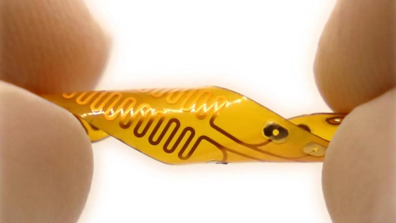
Dressings that simply cover wounds may soon seem archaic. An experimental new device reportedly speeds healing by 25%, and utilizes a computer-linked camera to determine when it should zap wounds with electricity or shoot medication into them. Known as a-Heal, the AI-enabled gadget is being developed by Prof. Marco Rolandi and colleagues at the University of California, Santa Cruz. Among other components, it incorporates a miniature fluorescence camera for imaging the wound, a ring of 12 LEDs for illumination, electrodes for electrically stimulating the wound site, plus reservoirs and bioelectronic actuators for storing and delivering liquid medication. The whole thing, along with a power source, is adhered to the skin overtop of the wound. Once the a-Heal has been set in place and activated, it proceeds to capture images of the wound once every two hours, and wirelessly transmits those images to a nearby computer. An AI agent on that computer analyzes the images, comparing the wound's current state of healing to where it ideally should be. If it's determined that the wound isn't healing fast enough, the agent prompts the a-Heal to either deliver an electric field to the wound site - thus enhancing cell migration toward wound closure - or to administer a dose of medication. In the case of a trial that was recently conducted on pigs over a 22-day period, that medication was a selective serotonin reuptake inhibitor known as fluoxetine, which increases wound tissue closure by decreasing inflammation. As a result of this combined approach, wounds on the pigs healed about 25% faster than those on a control group. It is hoped that the technology could be particularly useful in underserved regions where patients don't have ready access to modern medical facilities. "Our system takes all the cues from the body, and with external interventions, it optimizes the healing progress," says Rolandi. A paper on the research was recently published in the journal npj Biomedical Innovations.
Share
Share
Copy Link
Researchers at UC Santa Cruz have developed a-Heal, an AI-driven smart bandage that accelerates wound healing by 25%. This innovative device combines real-time imaging, machine learning, and bioelectronics to provide personalized wound treatment.

Introducing a-Heal: The AI-Powered Smart Bandage
Researchers at the University of California, Santa Cruz have developed a groundbreaking device called 'a-Heal' that promises to revolutionize wound treatment. This innovative smart bandage combines artificial intelligence, bioelectronics, and real-time imaging to accelerate wound healing by an impressive 25%
1
2
.How a-Heal Works
The a-Heal system consists of two main components: a wearable device and an AI-driven diagnostic and treatment algorithm called the 'ML Physician'
1
. The wearable device, which attaches to a commercially available bandage, includes:- A miniature camera that captures wound images every two hours
- LED illumination for clear imaging
- Bioelectronic actuators for drug delivery and electric field application
- A wireless transmission system for data communication
The ML Physician analyzes the wound images and determines the optimal treatment strategy based on the healing stage
2
.AI-Driven Personalized Treatment
The AI model employs a reinforcement learning approach, mimicking the diagnostic process used by human physicians. It uses an algorithm called 'Deep Mapper' to process wound images and quantify the healing stage compared to normal progression
3
.Based on this analysis, the system can administer two types of treatments:
- Topical application of fluoxetine, a selective serotonin reuptake inhibitor that reduces inflammation and promotes wound closure
- Application of an electric field to enhance cell migration towards wound closure
Related Stories

Promising Results and Future Implications
In preclinical studies conducted on pigs, wounds treated with a-Heal healed approximately 25% faster than those receiving standard care
4
. This significant improvement highlights the potential of a-Heal not only for accelerating acute wound healing but also for jump-starting the healing process in chronic wounds.The portable and wireless nature of a-Heal could make advanced wound therapy more accessible to patients in remote areas or those with limited mobility. As Professor Marco Rolandi, the lead researcher, explains, "Our system takes all the cues from the body, and with external interventions, it optimizes the healing progress"
2
.The Future of Wound Care
While a-Heal is still in the experimental stage, its success in preclinical trials suggests a promising future for AI-driven wound care. As the technology continues to develop, it could potentially transform the treatment of various types of wounds, from surgical incisions to chronic diabetic ulcers.
The integration of AI, bioelectronics, and real-time imaging in wound care represents a significant leap forward in medical technology. As research progresses, we may see similar smart devices applied to other areas of healthcare, ushering in a new era of personalized, AI-assisted medical treatments.
References
Summarized by
Navi
[2]
Related Stories
USC Researchers Develop AI-Powered Wireless Implant for Chronic Pain Management
25 Jun 2025•Science and Research
Mayo Clinic Develops AI Tool to Detect Surgical Site Infections from Patient Photos
08 Jul 2025•Health

Smart Insoles: Revolutionizing Sports Performance and Health Monitoring with AI-Powered Wearable Technology
27 Mar 2025•Technology

Recent Highlights
1
OpenAI Releases GPT-5.4, New AI Model Built for Agents and Professional Work
Technology

2
Anthropic sues Pentagon over supply chain risk label after refusing autonomous weapons use
Policy and Regulation

3
OpenAI secures $110 billion funding round as questions swirl around AI bubble and profitability
Business and Economy

Recent Highlights
Today's Top Stories


Your Daily Dose of Curated AI News
Don’t drown in AI news. We cut through the noise - filtering, ranking and summarizing the most important AI news, breakthroughs and research daily. Spend less time searching for the latest in AI and get straight to action.