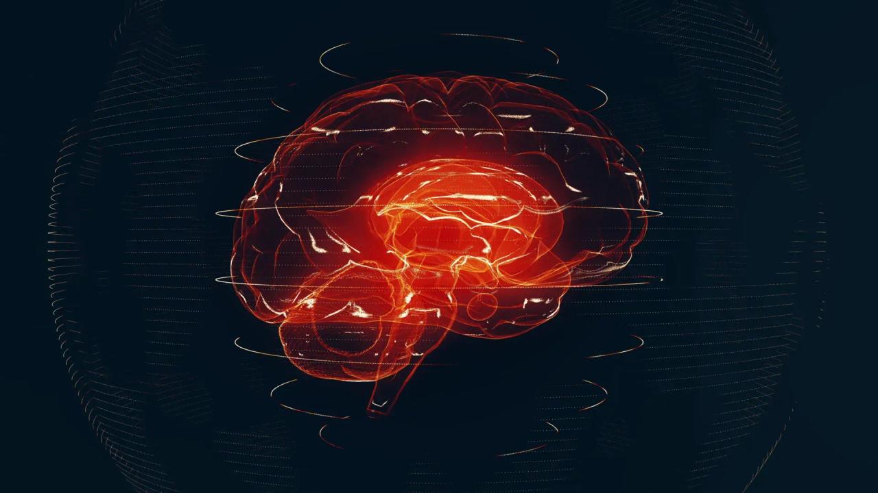
University of Michigan's Prima AI model reads brain MRI scans in seconds with 97.5% accuracy
4 Sources
4 Sources
[1]
Learning neuroimaging models from health system-scale data - Nature Biomedical Engineering
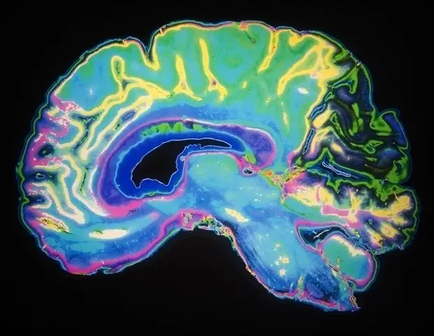
Neuroimaging is a ubiquitous tool for evaluating patients with neurological diseases. The global demand for magnetic resonance imaging (MRI) studies has risen steadily, placing substantial strain on health systems, prolonging turnaround times and intensifying physician burnout. These challenges disproportionately impact patients in low-resource and rural settings. Here we utilize data from a large academic health system to develop Prima, an AI foundation model for neuroimaging that supports real-world, clinical MRI studies as input. Trained on over 220,000 MRI studies, Prima uses a hierarchical vision architecture that provides general and transferable MRI features. Prima was tested in a 1-year health system-wide study that included 29,431 MRI studies. Across 52 radiologic diagnoses from major neurologic disorders, Prima achieved a mean diagnostic area under the curve (AUC) of 92.0%, outperforming other state-of-the-art general and medical AI models. Prima offers explainable differential diagnoses, worklist priority for radiologists and clinical referral recommendations. Prima demonstrates algorithmic fairness across sensitive groups. These findings highlight the transformative potential of health system-scale AI training and Prima's role in advancing AI-driven healthcare.
[2]
AI reads brain MRIs in seconds and flags emergencies
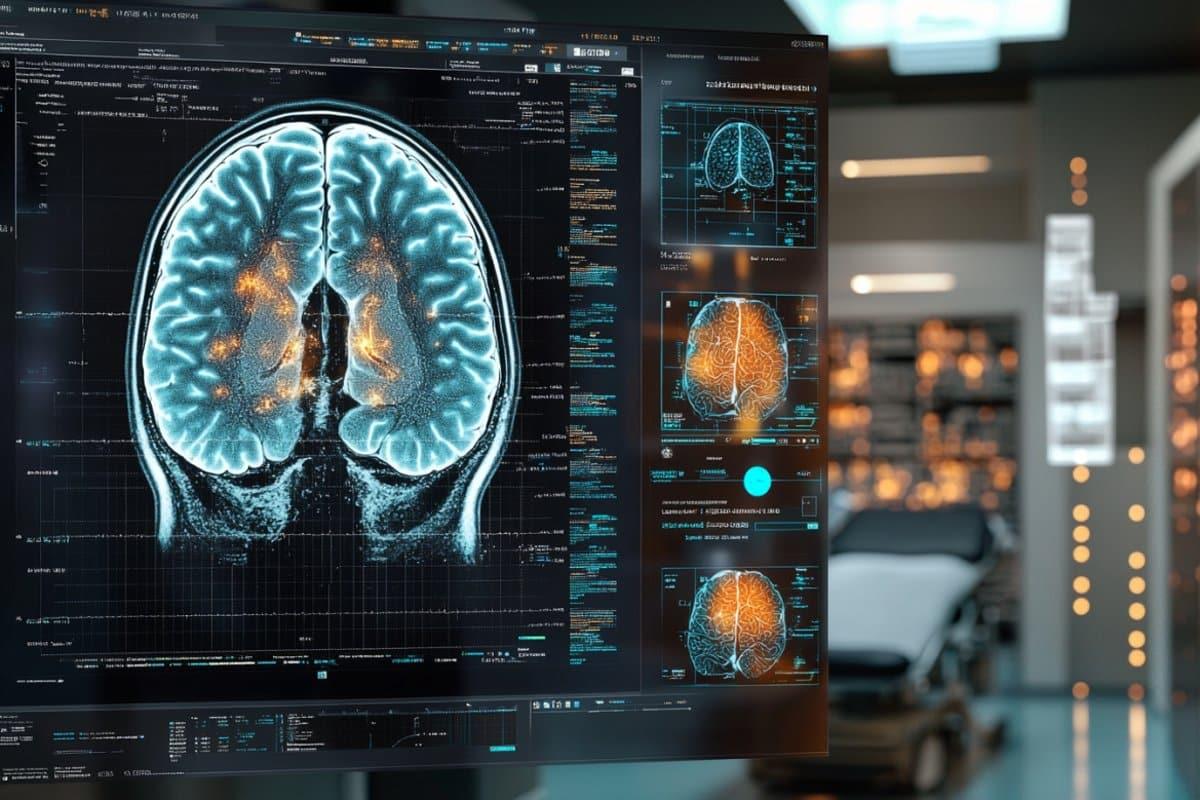
A newly developed artificial intelligence system from the University of Michigan can analyze brain MRI scans and deliver a diagnosis in a matter of seconds, according to a new study. The model identified neurological conditions with accuracy reaching 97.5% and was also able to assess how urgently patients needed medical care. Researchers say this first-of-its-kind technology has the potential to reshape how brain imaging is handled across health systems in the United States. The findings were published in Nature Biomedical Engineering. "As the global demand for MRI rises and places significant strain our physicians and health systems, our AI model has potential to reduce burden by improving diagnosis and treatment with fast, accurate information," said senior author Todd Hollon, M.D., a neurosurgeon at University of Michigan Health and assistant professor of neurosurgery at U-M Medical School. Testing the Prima AI System Hollon named the new technology Prima. Over a one-year period, his research team evaluated the system using more than 30,000 MRI studies. Across more than 50 different radiologic diagnoses involving major neurological disorders, Prima delivered stronger diagnostic performance than other advanced AI models. In addition to identifying disease, the system also proved capable of determining which cases required higher priority. Certain neurological conditions, including strokes and brain hemorrhages, demand immediate medical attention. Hollon said that in these situations, Prima can automatically alert health care providers so action can be taken quickly. The system was designed to notify the most appropriate subspecialist, such as a stroke neurologist or neurosurgeon. Feedback becomes available immediately after a patient completes imaging. "Accuracy is paramount when reading a brain MRI, but quick turnaround times are critical for timely diagnosis and improved outcomes," said Yiwei Lyu, M.S., co-first author and postdoctoral fellow of Computer Science and Engineering at U-M. "At key steps in the process, our results show how Prima can improve workflows and streamline clinical care without abandoning accuracy." What Is Prima? Prima is classified as a vision language model (VLM), a type of artificial intelligence that can process images, video, and text together in real time. While artificial intelligence has been applied to MRI analysis before, researchers say Prima takes a different approach. Earlier models were typically trained on carefully selected subsets of MRI data and designed to perform narrow tasks, such as identifying lesions or estimating dementia risk. Prima was trained on a much broader dataset. Hollon's team used every available MRI collected since radiology records were digitized at University of Michigan Health. This included more than 200,000 MRI studies and 5.6 million imaging sequences. The model also incorporated patients' clinical histories and the reasons physicians ordered each imaging study. "Prima works like a radiologist by integrating information regarding the patient's medical history and imaging data to produce a comprehensive understanding of their health," said co-first author Samir Harake, a data scientist in Hollon's Machine Learning in Neurosurgery Lab. "This enables better performance across a broad range of prediction tasks." Addressing MRI Delays and Radiology Shortages Each year, millions of MRI scans are performed worldwide, many of them focused on neurological disease. Researchers say the demand for these scans is growing faster than the availability of neuroradiology services. This imbalance has contributed to staffing shortages, diagnostic delays, and errors. Depending on where a patient receives a scan, results may take days or even longer to return. "Whether you are receiving a scan at a larger health system that is facing increasing volume or a rural hospital with limited resources, innovative technologies are needed to improve access to radiology services," said Vikas Gulani, M.D. Ph.D., co-author and chair of the Department of Radiology at U-M Health. "Our teams at University of Michigan have collaborated to develop a cutting-edge solution to this problem with tremendous, scalable potential." The Future of AI in Medical Imaging Although Prima performed strongly, researchers emphasize that the work is still in an early evaluation phase. Future research will focus on incorporating more detailed patient information and electronic medical record data to further improve diagnostic accuracy. This approach mirrors how radiologists and physicians interpret MRIs and other imaging studies in real clinical settings. While artificial intelligence is already used in health care, most existing systems are limited to narrowly defined tasks. Hollon describes Prima as "ChatGPT for medical imaging," noting that similar technology could eventually be adapted for other imaging types, including mammograms, chest X-rays and ultrasounds. "Like the way AI tools can help draft an email or provide recommendations, Prima aims to be a co-pilot for interpreting medical imaging studies," Hollon said. "We believe that Prima exemplifies the transformative potential of integrating health systems and AI-driven models to improve health care through innovation." Additional authors: Asadur Chowdury, M.S., Soumyanil Banerjee, M.S., Rachel Gologorsky, Shixuan Liu, Anna-Katharina Meissner, M.D., Akshay Rao, Chenhui Zhao, Akhil Kondepudi, Cheng Jiang, Xinhai Hou, Rushikesh S. Joshi, M.D., Volker Neuschmelting, M.D., Ashok Srinivasan, M.D., Dawn Kleindorfer, M.D., Brian Athey, Ph.D., Aditya Pandey, M.D., and Honglak Lee, Ph.D., all of University of Michigan. Funding/disclosures: This work was supported in part by the National Institute of Neurological Disorders and Stroke (K12NS080223) of the National Institutes of Health. The content is solely the responsibility of the authors and does not necessarily represent the official views of the NIH. This work was also supported by the Chan Zuckerberg Initiative (CZI), Frankel Institute for Heart and Brain Health, the Mark Trauner Brain Research Fund, the Zenkel Family Foundation, Ian's Friends Foundation and the UM Precision Health Investigators Awards grant program.
[3]
AI Model Can Read and Diagnose a Brain MRI in Seconds | Newswise
Newswise -- An AI-powered model developed at University of Michigan can read a brain MRI and diagnose a person in seconds, a study suggests. The model detected neurological conditions with up to 97.5% accuracy and predicted how urgently a patient required treatment. Researchers say the first-of-its-kind technology could transform neuroimaging at health systems across the United States. The results are published in Nature Biomedical Engineering. "As the global demand for MRI rises and places significant strain our physicians and health systems, our AI model has potential to reduce burden by improving diagnosis and treatment with fast, accurate information," said senior author Todd Hollon, M.D., a neurosurgeon at University of Michigan Health and assistant professor of neurosurgery at U-M Medical School. Hollon calls the invention Prima. He and his research team tested the technology on more than 30,000 MRI studies over the course of a year. Across more than 50 radiologic diagnoses from major neurological disorders, Prima outperformed other state-of-the-art AI models on diagnostic performance. The model also succeeded in determining which cases should take higher priority. Some neurological conditions, such as brain hemorrhages or strokes, require immediate medical attention. In such cases, Prima can automatically alert providers so rapid action can be taken, Hollon says. Researchers designed the model to recommend which subspecialty provider should be alerted, such as a stroke neurologist or neurosurgeon, with feedback available immediately after a patient completes imaging. "Accuracy is paramount when reading a brain MRI, but quick turnaround times are critical for timely diagnosis and improved outcomes," said Yiwei Lyu, M.S., co-first author and postdoctoral fellow of Computer Science and Engineering at U-M. "At key steps in the process, our results show how Prima can improve workflows and streamline clinical care without abandoning accuracy." What is Prima? Prima is a vision language model (VLM), an AI system that can simultaneously process video, images and text in real time. It's not the first attempt to apply AI to MRI and other forms of neuroimaging, but the approach is unique. Past models rely on manually curated subsets of MRI data to achieve specific tasks, such detecting lesions or predicting dementia risk. When designing Prima, Hollon's team trained the system on every MRI -- over 200,000 studies and 5.6 million sequences -- taken since radiology digitization began University of Michigan Health decades ago. Researchers also input patients' clinical histories and the physicians' reasons for ordering medical imaging study into the model. "Prima works like a radiologist by integrating information regarding the patient's medical history and imaging data to produce a comprehensive understanding of their health," said co-first author Samir Harake, a data scientist in Hollon's Machine Learning in Neurosurgery Lab. "This enables better performance across a broad range of prediction tasks." Millions of MRI studies are performed globally each year, with a significant portion focused on neurological diseases. This demand, researchers say, outpaces the availability of neuroradiology services and leads to significant challenges, including workforce shortages and diagnostic errors. Depending on where you get a scan, it can take days, or even longer, to get a result. "Whether you are receiving a scan at a larger health system that is facing increasing volume or a rural hospital with limited resources, innovative technologies are needed to improve access to radiology services," said Vikas Gulani, M.D. Ph.D., co-author and chair of the Department of Radiology at U-M Health. "Our teams at University of Michigan have collaborated to develop a cutting-edge solution to this problem with tremendous, scalable potential." The future of AI and imaging While Prima performed well, the research is in its initial stage of evaluation. The research team's future work will explore integrating more detailed patient information and electronic medical record data for more accurate diagnosis. This strategy closely emulates how radiologists and physicians interpret MRIs and other radiology studies. Health care providers, systems and policymakers are still determining how to appropriately integrate artificial intelligence into practice, yet most systems currently used are for narrow medical tasks. What Hollon describes as "ChatGPT for medical imaging" has broader potential -- and could one day be adapted for other imaging modalities, such as mammograms, chest X-rays and ultrasounds. "Like the way AI tools can help draft an email or provide recommendations, Prima aims to be a co-pilot for interpreting medical imaging studies," Hollon said. "We believe that Prima exemplifies the transformative potential of integrating health systems and AI-driven models to improve health care through innovation." Additional authors: Asadur Chowdury, M.S., Soumyanil Banerjee, M.S., Rachel Gologorsky, Shixuan Liu, Anna-Katharina Meissner, M.D., Akshay Rao, Chenhui Zhao, Akhil Kondepudi, Cheng Jiang, Xinhai Hou, Rushikesh S. Joshi, M.D., Volker Neuschmelting, M.D., Ashok Srinivasan, M.D., Dawn Kleindorfer, M.D., Brian Athey, Ph.D., Aditya Pandey, M.D., and Honglak Lee, Ph.D., all of University of Michigan. Funding/disclosures: This work was supported in part by the National Institute of Neurological Disorders and Stroke (K12NS080223) of the National Institutes of Health. The content is solely the responsibility of the authors and does not necessarily represent the official views of the NIH. This work was also supported by the Chan Zuckerberg Initiative (CZI), Frankel Institute for Heart and Brain Health, the Mark Trauner Brain Research Fund, the Zenkel Family Foundation, Ian's Friends Foundation and the UM Precision Health Investigators Awards grant program. Michigan Research Core(s): UM Advanced Research Computing Paper cited: "Learning neuroimaging models from health system-scale data," Nature Biomedical Engineering. DOI: 10.1038/s41551-025-01608-0
[4]
Why doctors believe this new AI system will end MRI wait times
A new artificial intelligence system, named Prima, developed at the University of Michigan, can diagnose neurological conditions from brain MRI scans within seconds. The system achieved 97.5% accuracy in identifying neurological conditions and could also assess the urgency of medical care required by patients. This technology aims to redefine brain imaging practices within U.S. health systems. The findings regarding Prima were published in the journal Nature Biomedical Engineering. Dr. Todd Hollon, a neurosurgeon at University of Michigan Health and assistant professor of neurosurgery at U-M Medical School, led the research team. Dr. Hollon is also a senior author of the study. Over a one-year period, Dr. Hollon's team evaluated Prima using more than 30,000 MRI studies. The system demonstrated superior diagnostic performance compared to other advanced AI models across over 50 different radiologic diagnoses of major neurological disorders. Prima can identify conditions such as strokes and brain hemorrhages, which require immediate medical attention. In these instances, the system can automatically alert healthcare providers, including subspecialists like stroke neurologists or neurosurgeons, for rapid intervention. Feedback becomes available immediately after a patient completes imaging. Yiwei Lyu, a co-first author and postdoctoral fellow of Computer Science and Engineering at U-M, noted that timely diagnosis is critical for improved patient outcomes. Prima is classified as a vision language model (VLM), capable of processing images, video, and text in real time. Unlike earlier AI models trained on limited MRI subsets for narrow tasks, Prima was trained on a broad dataset encompassing over 200,000 MRI studies and 5.6 million imaging sequences collected since radiology records were digitized at University of Michigan Health. The model also incorporated patients' clinical histories and the reasons physicians ordered each imaging study. Samir Harake, a co-first author and data scientist in Hollon's Machine Learning in Neurosurgery Lab, explained that Prima integrates patient medical history and imaging data, similar to a radiologist, to provide a comprehensive understanding of a patient's health. Millions of MRI scans are performed globally each year, with a rising demand for neuroradiology services. This increasing demand, coupled with shortages in neuroradiology staff, contributes to diagnostic delays and errors. Patients often experience wait times of days or longer for MRI results. Dr. Vikas Gulani, co-author and chair of the Department of Radiology at U-M Health, stated that innovative technologies are necessary to improve access to radiology services in both large health systems and rural hospitals with limited resources. Researchers consider Prima to be in an early evaluation phase. Future research will focus on integrating more detailed patient information and electronic medical record data to enhance diagnostic accuracy. This approach mirrors how radiologists and physicians interpret medical imaging studies in clinical settings. Dr. Hollon compares Prima to "ChatGPT for medical imaging," suggesting similar technology could be adapted for other imaging types, including mammograms, chest X-rays, and ultrasounds.
Share
Share
Copy Link
Researchers at University of Michigan developed Prima, an AI model that analyzes brain MRI scans and delivers diagnoses in seconds with up to 97.5% accuracy. Trained on over 220,000 MRI studies, Prima can prioritize emergency cases like strokes and brain hemorrhages, automatically alerting specialists. The system aims to address growing radiology shortages and reduce diagnostic delays across health systems.
Prima AI Model Transforms Brain MRI Diagnosis Speed
Researchers at the University of Michigan have developed Prima, an AI model that can analyze brain MRI scans and deliver diagnoses in seconds, achieving up to 97.5% accuracy in detecting neurological conditions
1
. Published in Nature Biomedical Engineering, this breakthrough technology addresses a critical gap in healthcare as global demand for MRI studies continues to strain health systems and extend turnaround times2
. Led by Todd Hollon, a neurosurgeon at University of Michigan Health, the research team tested Prima on 29,431 MRI studies over a one-year period, demonstrating its potential to reshape how brain imaging is handled across the United States3
.
Source: ScienceDaily
Trained on MRI Studies at Unprecedented Scale
Unlike previous AI systems trained on carefully curated subsets of MRI data for narrow tasks, Prima was trained on over 220,000 MRI studies and 5.6 million imaging sequences collected since radiology digitization began at University of Michigan Health decades ago
1
. The system incorporates patient clinical histories and physicians' reasons for ordering each imaging study, enabling it to function like a radiologist by integrating multiple data sources3
. Samir Harake, co-first author and data scientist in Hollon's Machine Learning in Neurosurgery Lab, explains that this comprehensive approach produces a better understanding of patient health and enables superior diagnostic performance across a broad range of prediction tasks2
.High Diagnostic Accuracy Across Neurological Conditions
Across 52 radiologic diagnoses from major neurological disorders, Prima achieved a mean diagnostic area under the curve of 92.0%, outperforming other state-of-the-art general and medical AI models
1
. The system demonstrated algorithmic fairness across sensitive groups and offers explainable differential diagnoses to clinicians1
. Yiwei Lyu, co-first author and postdoctoral fellow of Computer Science and Engineering at U-M, emphasizes that while accuracy is paramount when reading a brain MRI, quick turnaround times are critical for timely diagnosis and improved outcomes4
.Vision Language Model Architecture Enables Real-Time Analysis
Prima functions as a vision language model (VLM), an AI system that can simultaneously process video, images, and text in real time
3
. This architecture allows Prima to integrate diverse data types and deliver comprehensive diagnostic insights immediately after a patient completes imaging2
. The foundation model uses a hierarchical vision architecture that provides general and transferable MRI features, enabling it to handle real-world clinical MRI studies as input1
.Prioritize Emergency Cases and Reduce MRI Wait Times
Prima can automatically identify conditions requiring immediate medical attention, such as strokes and brain hemorrhages, and alert the appropriate subspecialist—whether a stroke neurologist or neurosurgeon
2
. The system provides worklist prioritization for radiologists and clinical referral recommendations, with feedback available immediately after imaging completion1
. This capability addresses a pressing need as millions of MRI scans are performed globally each year, with demand outpacing the availability of neuroradiology services and contributing to diagnostic delays that can extend for days or longer depending on location3
.Related Stories


Address Radiology Shortages Across Health Systems
Vikas Gulani, co-author and chair of the Department of Radiology at U-M Health, notes that innovative technologies are essential to improve access to radiology services whether patients receive scans at larger health systems facing increasing volume or rural hospitals with limited resources
2
. The imbalance between growing MRI demand and available neuroradiology services has contributed to staffing shortages, diagnostic delays, and errors—challenges that disproportionately impact patients in low-resource and rural settings1
. Hollon describes Prima as "ChatGPT for medical imaging," suggesting the technology could eventually be adapted for other imaging modalities including mammograms, chest X-rays, and ultrasounds4
.Future Development to Diagnose Neurological Conditions More Accurately
While Prima performed strongly in initial testing, researchers emphasize the work remains in an early evaluation phase
2
. Future research will focus on integrating more detailed patient information and electronic medical record data to further improve diagnostic accuracy, mirroring how radiologists and physicians interpret MRIs in real clinical settings3
. Hollon believes Prima exemplifies the transformative potential of integrating health systems and AI-driven models to improve healthcare through innovation, positioning the system as a co-pilot for interpreting medical imaging studies rather than a replacement for human expertise3
.References
Summarized by
Navi
[1]
[2]
Related Stories
Recent Highlights
1
Tennessee Teens Sue Elon Musk's xAI Over Grok AI-Generated Child Abuse Images
Policy and Regulation

2
Supermicro Co-Founder Indicted in $2.5 Billion Nvidia AI Chip Smuggling Scheme to China
Policy and Regulation
3
Val Kilmer to appear posthumously in As Deep as the Grave through AI-generated performance
Entertainment and Society

Recent Highlights
Today's Top Stories


Your Daily Dose of Curated AI News
Don’t drown in AI news. We cut through the noise - filtering, ranking and summarizing the most important AI news, breakthroughs and research daily. Spend less time searching for the latest in AI and get straight to action.