AI-Powered 'Segment Anything for Microscopy' Revolutionizes Cell Analysis
3 Sources
3 Sources
[1]
Automatic cell analysis with the help of artificial intelligence
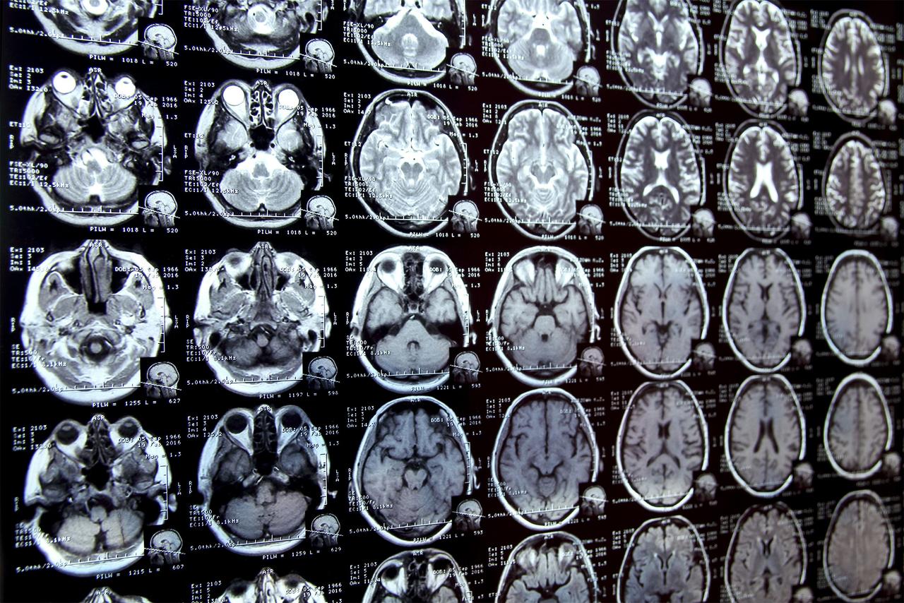
Identifying and delineating cell structures in microscopy images is crucial for understanding the complex processes of life. This task is called "segmentation" and it enables a range of applications, such as analysing the reaction of cells to drug treatments, or comparing cell structures in different genotypes. It was already possible to carry out automatic segmentation of those biological structures but the dedicated methods only worked in specific conditions and adapting them to new conditions was costly. An international research team led by Göttingen University has now developed a method by retraining the existing AI-based software Segment Anything on over 17,000 microscopy images with over 2 million structures annotated by hand. Their new model is called Segment Anything for Microscopy and it can precisely segment images of tissues, cells and similar structures in a wide range of settings. To make it available to researchers and medical doctors, they have also created μSAM, a user-friendly software to "segment anything" in microscopy images. Their work was published in Nature Methods. To adapt the existing software to microscopy, the research team first evaluated it on a large set of open-source data, which showed the model's potential for microscopy segmentation. To improve quality, the team retrained it on a large microscopy dataset. This dramatically improved the model's performance for the segmentation of cells, nuclei and tiny structures in cells known as organelles. The team then created their software, μSAM, which enables researchers and medical doctors to analyse images without the need to first manually paint structures or train a specific AI model. The software is already in wide use internationally, for example to analyse nerve cells in the ear as part of a project on hearing restoration, to segment artificial tumour cells for cancer research, or to analyse electron microscopy images of volcanic rocks. "Analysing cells or other structures is one of the most challenging tasks for researchers working in microscopy and is an important task for both basic research in biology and medical diagnostics," says Junior Professor Constantin Pape at Göttingen University's Institute of Computer Science. "My group specializes in building tools to automate such tasks and we often get asked by researchers to help. Before the development of Segment Anything for Microscopy, we had to ask them to first annotate a lot of structures by hand - a difficult and time-consuming task. μSAM has changed this! Tasks that used to take weeks of painstaking manual effort can be automated in a few hours, because the model can segment any kind of biological structure with a few clicks and can then be further improved to automate the task with our tool. This enables many new applications, and we have already used it in a wide range of projects, ranging from basic cell biology to developing tools for treatment recommendation in cancer therapies."
[2]
Automatic cell analysis: AI-powered software 'segments anything' in microscopy images
Identifying and delineating cell structures in microscopy images is crucial for understanding the complex processes of life. This task is called "segmentation" and it enables a range of applications, such as analyzing the reaction of cells to drug treatments, or comparing cell structures in different genotypes. It was already possible to carry out automatic segmentation of those biological structures, but the dedicated methods only worked in specific conditions and adapting them to new conditions was costly. An international research team led by Göttingen University has now developed a method for retraining the existing AI-based software Segment Anything on over 17,000 microscopy images with over 2 million structures annotated by hand. The new model is called Segment Anything for Microscopy and it can precisely segment images of tissues, cells and similar structures in a wide range of settings. To make it available to researchers and medical doctors, they have also created μSAM, user-friendly software to "segment anything" in microscopy images. The work is published in Nature Methods. To adapt the existing software to microscopy, the research team first evaluated it on a large set of open-source data, which showed the model's potential for microscopy segmentation. To improve quality, the team retrained it on a large microscopy dataset. This dramatically improved the model's performance for the segmentation of cells, nuclei and tiny structures in cells known as organelles. The team then created their software, μSAM, which enables researchers and medical doctors to analyze images without the need to first manually paint structures or train a specific AI model. The software is already in wide use internationally; for example, to analyze nerve cells in the ear as part of a project on hearing restoration, to segment artificial tumor cells for cancer research, or to analyze electron microscopy images of volcanic rocks. "Analyzing cells or other structures is one of the most challenging tasks for researchers working in microscopy and is an important task for both basic research in biology and medical diagnostics," says Junior Professor Constantin Pape at Göttingen University's Institute of Computer Science. "My group specializes in building tools to automate such tasks and we often get asked by researchers to help. Before the development of Segment Anything for Microscopy, we had to ask them to first annotate a lot of structures by hand -- a difficult and time-consuming task. μSAM has changed this. Tasks that used to take weeks of painstaking manual effort can be automated in a few hours, because the model can segment any kind of biological structure with a few clicks and can then be further improved to automate the task with our tool. "This enables many new applications, and we have already used it in a wide range of projects, ranging from basic cell biology to developing tools for treatment recommendation in cancer therapies."
[3]
New AI model revolutionizes cell structure segmentation in microscopy
University of GöttingenFeb 26 2025 Identifying and delineating cell structures in microscopy images is crucial for understanding the complex processes of life. This task is called "segmentation" and it enables a range of applications, such as analyzing the reaction of cells to drug treatments, or comparing cell structures in different genotypes. It was already possible to carry out automatic segmentation of those biological structures but the dedicated methods only worked in specific conditions and adapting them to new conditions was costly. An international research team led by Göttingen University has now developed a method by retraining the existing AI-based software Segment Anything on over 17,000 microscopy images with over 2 million structures annotated by hand. Their new model is called Segment Anything for Microscopy and it can precisely segment images of tissues, cells and similar structures in a wide range of settings. To make it available to researchers and medical doctors, they have also created μSAM, a user-friendly software to "segment anything" in microscopy images. Their work was published in Nature Methods. To adapt the existing software to microscopy, the research team first evaluated it on a large set of open-source data, which showed the model's potential for microscopy segmentation. To improve quality, the team retrained it on a large microscopy dataset. This dramatically improved the model's performance for the segmentation of cells, nuclei and tiny structures in cells known as organelles. The team then created their software, μSAM, which enables researchers and medical doctors to analyze images without the need to first manually paint structures or train a specific AI model. The software is already in wide use internationally, for example to analyze nerve cells in the ear as part of a project on hearing restoration, to segment artificial tumor cells for cancer research, or to analyze electron microscopy images of volcanic rocks. Analyzing cells or other structures is one of the most challenging tasks for researchers working in microscopy and is an important task for both basic research in biology and medical diagnostics. My group specializes in building tools to automate such tasks and we often get asked by researchers to help. Before the development of Segment Anything for Microscopy, we had to ask them to first annotate a lot of structures by hand - a difficult and time-consuming task. μSAM has changed this! Tasks that used to take weeks of painstaking manual effort can be automated in a few hours, because the model can segment any kind of biological structure with a few clicks and can then be further improved to automate the task with our tool. This enables many new applications, and we have already used it in a wide range of projects, ranging from basic cell biology to developing tools for treatment recommendation in cancer therapies." Junior Professor Constantin Pape at Göttingen University's Institute of Computer Science University of Göttingen Journal reference: Archit, A. (2025). Segment Anything for Microscopy. Nature Methods. doi.org/10.1038/s41592-024-02580-4.
Share
Share
Copy Link
Researchers at Göttingen University have developed an AI model that dramatically improves microscopy image segmentation, potentially accelerating biological research and medical diagnostics.

AI Breakthrough in Microscopy Image Analysis
An international research team led by Göttingen University has developed a groundbreaking AI model called "Segment Anything for Microscopy" (SAM), which promises to revolutionize the analysis of cell structures in microscopy images. This innovation, published in Nature Methods, addresses a critical challenge in biological research and medical diagnostics
1
.The Power of Segmentation
Segmentation, the process of identifying and delineating cell structures in microscopy images, is crucial for understanding complex biological processes. It enables researchers to analyze cellular responses to drug treatments and compare cell structures across different genotypes. While automatic segmentation methods existed previously, they were limited to specific conditions and costly to adapt
2
.Development of Segment Anything for Microscopy
The research team adapted the existing AI-based software "Segment Anything" by retraining it on an extensive dataset:
- Over 17,000 microscopy images
- More than 2 million hand-annotated structures
This retraining process dramatically improved the model's performance in segmenting cells, nuclei, and organelles across a wide range of settings
3
.μSAM: User-Friendly Interface
To make this powerful tool accessible to researchers and medical professionals, the team developed μSAM, a user-friendly software interface. μSAM allows users to "segment anything" in microscopy images without the need for manual structure painting or specific AI model training
1
.Wide-Ranging Applications
The software is already being used internationally for various applications:
- Analyzing nerve cells in the ear for hearing restoration projects
- Segmenting artificial tumor cells in cancer research
- Examining electron microscopy images of volcanic rocks
Related Stories


Impact on Research Efficiency
Junior Professor Constantin Pape from Göttingen University's Institute of Computer Science highlighted the significant time-saving aspect of the new tool:
"Tasks that used to take weeks of painstaking manual effort can be automated in a few hours, because the model can segment any kind of biological structure with a few clicks and can then be further improved to automate the task with our tool."
2
Future Prospects
The development of Segment Anything for Microscopy opens up new possibilities in various fields:
- Basic cell biology research
- Development of tools for treatment recommendations in cancer therapies
- Potential applications in other areas of medical diagnostics and biological research
As the tool continues to be adopted and refined, it has the potential to significantly accelerate scientific discoveries and improve medical diagnoses across a wide range of disciplines.
References
Summarized by
Navi
[1]
Related Stories
Recent Highlights


Recent Highlights
Today's Top Stories


Your Daily Dose of Curated AI News
Don’t drown in AI news. We cut through the noise - filtering, ranking and summarizing the most important AI news, breakthroughs and research daily. Spend less time searching for the latest in AI and get straight to action.