MIT's AI System Revolutionizes Medical Image Segmentation for Clinical Research


3 Sources
[1]
New AI system could accelerate clinical research
Annotating regions of interest in medical images, a process known as segmentation, is often one of the first steps clinical researchers take when running a new study involving biomedical images. For instance, to determine how the size of the brain's hippocampus changes as patients age, the
[2]
New AI system streamlines segmentation of biomedical images
Massachusetts Institute of TechnologySep 26 2025 Annotating regions of interest in medical images, a process known as segmentation, is often one of the first steps clinical researchers take when running a new study involving biomedical images. For instance, to determine how the size of the
[3]
AI system for rapid annotation of medical images could accelerate clinical research
Annotating regions of interest in medical images, a process known as segmentation, is often one of the first steps clinical researchers take when running a new study involving biomedical images. For instance, to determine how the size of the brain's hippocampus changes as patients age, the
Share
Copy Link
MIT researchers have developed an AI-based system called MultiverSeg that streamlines the process of segmenting biomedical images. This innovative tool could significantly accelerate clinical research and reduce costs in medical studies.
MIT Researchers Develop Groundbreaking AI for Medical Image Segmentation
MIT researchers have unveiled a cutting-edge artificial intelligence system that promises to revolutionize the field of medical image segmentation, potentially accelerating clinical research and reducing costs in medical studies
1
.The Challenge of Medical Image Segmentation
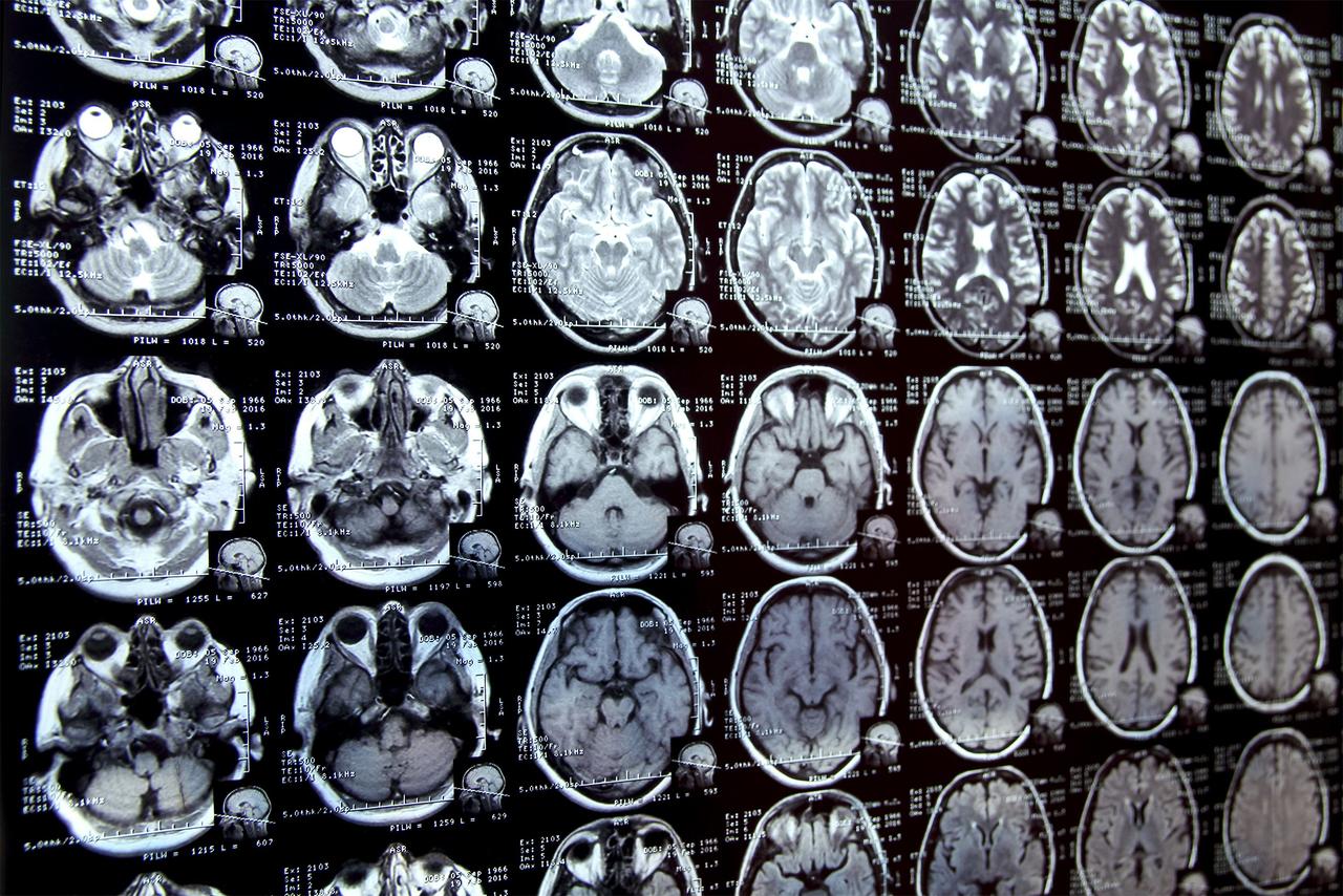
Segmentation, the process of annotating regions of interest in medical images, is a crucial first step in many clinical research studies. For example, when studying how the brain's hippocampus changes with age, researchers must outline this structure in numerous brain scans. This manual process can be extremely time-consuming, especially for complex structures
2
.
Source: MIT
Introducing MultiverSeg: A Game-Changing AI Solution
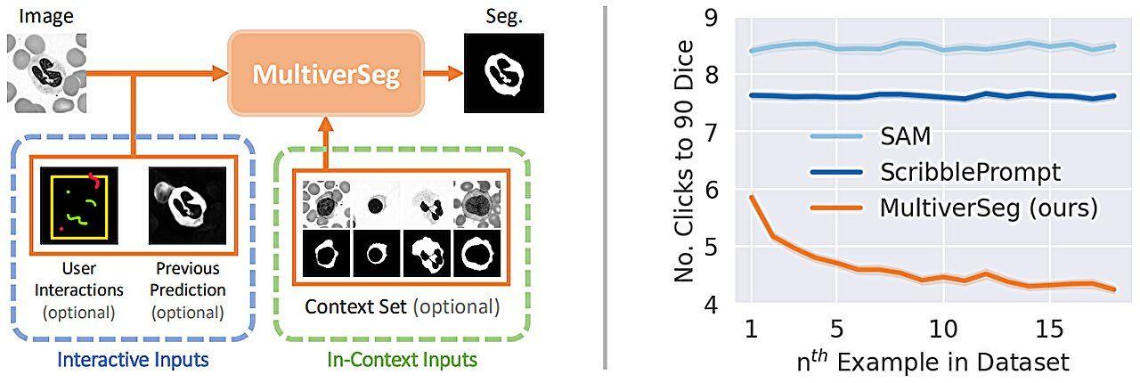
To address this challenge, MIT researchers have developed MultiverSeg, an AI-based system that enables rapid segmentation of biomedical imaging datasets. The system allows researchers to segment images by clicking, scribbling, and drawing boxes, with the AI model predicting the segmentation based on these interactions .
Key Features of MultiverSeg
-
Adaptive Learning: As users mark additional images, the system requires fewer interactions, eventually segmenting new images accurately without user input
1
. -
Context-Aware Architecture: The model's architecture is designed to use information from previously segmented images to make new predictions, improving accuracy over time
2
. -
Flexibility: MultiverSeg can work with context sets of any size, making it adaptable to various applications .
-
User-Friendly: The system doesn't require pre-segmented datasets for training or machine learning expertise, making it accessible to a wide range of researchers
1
.
Source: Medical Xpress
Related Stories




Potential Impact on Clinical Research
MultiverSeg has the potential to significantly accelerate studies of new treatment methods and reduce the cost of clinical trials and medical research. It could also improve the efficiency of clinical applications, such as radiation treatment planning
2
.Hallee Wong, the lead author of the study, emphasizes the system's potential: "Many scientists might only have time to segment a few images per day for their research because manual image segmentation is so time-consuming. Our hope is that this system will enable new science by allowing clinical researchers to conduct studies they were prohibited from doing before because of the lack of an efficient tool" .
Future Prospects and Ongoing Research
The research team, including Jose Javier Gonzalez Ortiz, John Guttag, and Adrian Dalca, will present their findings at the upcoming International Conference on Computer Vision. As MultiverSeg continues to develop, it may open new avenues for medical research and improve the efficiency of clinical applications, potentially transforming the landscape of biomedical imaging analysis
1
.References
Summarized by

Navi
[2]
Related Stories
Recent Highlights
1
Microsoft and Nvidia Launch Surface Laptop Ultra: A $2,599 AI-Ready PC With RTX Spark Chip
Technology

2
Trump Consulted Grok AI Before Ordering Venezuela Invasion and Capture of Nicolás Maduro
Policy and Regulation

3
Scientists Build AI Model That Reconstructs Images From Brain Scans in Record Time
Science and Research
Recent Highlights
Today's Top Stories






Don’t drown in AI news. We cut through the noise - filtering, ranking and summarizing the most important AI news, breakthroughs and research daily. Follow topics that matter to you and stay ahead.


