AI Tools Enhance Detection of Immune Structures in Melanoma, Improving Survival Predictions
2 Sources
2 Sources
[1]
AI tools improve detection of key immune structures in melanoma
ECOG-ACRIN Cancer Research GroupApr 24 2025 Researchers from the ECOG-ACRIN Cancer Research Group (ECOG-ACRIN) have applied AI-driven processes for detecting tertiary lymphoid structures (TLS) in thousands of digital images of melanoma tumor tissue, significantly enhancing TLS identification and survival predictions for operable stage III/IV patients. The presence of TLS, a key biomarker for better prognosis and improved survival, is not yet a standard part of patients' pathology reports, and manual detection is labor-intensive and can be variable. Lead investigators Ahmad A. Tarhini, MD, PhD, and Xuefeng Wang, PhD, will present the new approach at the American Association for Cancer Research 2025 Annual Meeting in Chicago. Our efforts reveal the potential of open-source AI tools to transform how we predict survival and immunotherapy benefits by detecting critical immune structures like TLS with unprecedented ease and accuracy." Dr. Ahmad A. Tarhini, professor and senior member, cutaneous oncology and immunology, at the Moffitt Cancer Center and Research Institute in Tampa, Florida The study retrospectively analyzed thousands of archived digital images coupled with corresponding RNA sequencing data from 376 patients with advanced, high-risk melanoma, linking TLS presence to significantly better overall survival. The cohort had participated in a landmark US cooperative group trial led by ECOG-ACRIN called E1609 that tested immune check point blockade and cytokine therapy in high-risk melanoma (Tarhini A. J Clin Oncol. February 2020). This analysis found TLS present in 55% of the E1609 cohort and predicted significantly better overall survival than those without TLS (36.23% vs. 29.59% at 5 years), especially in those with more than one TLS (38.04% in >1 TLS vs. 28.65%). TLS density was also significantly prognostic for overall survival (37.77% vs. 28.72% at 5 years for median cutoff). Survival also varied by AJCC stage group, age, sex, treatment type, and tumor ulceration, as shown in AACR Abstract 3358. "These findings highlight the potential for AI-driven approaches to standardize TLS assessment using low-cost H&E-stained images, with the potential to improve prognostication and stratification within AJCC, and warrant further investigation," said Dr. Tarhini. Researchers first applied HookNet-TLS, an open-source deep learning algorithm, to measure TLS and germinal centers (GC) within the E1609 digitized H&E-stained slides. After reviewing the initial results, they retrained the model for better accuracy. They evaluated the prognostic value of TLS scores by correlating the presence of TLS and GC found in the digitized images with normalized TLS counts. Next, the researchers applied Gigapth Whole-Slide Foundation Model for Digital Pathology feature extraction and investigated the potential in TLS detection in this cohort. Gigapth allowed for enhanced visualization of H&E image tiles through the generation of principal component analysis (PCA). "Utilizing Gigapth Foundation Model, the generated PCA visualizations appear promising in enhancing TLS and GC detection. These are undergoing further fine-tuning, and the final results will be shared at a future meeting," said Dr. Wang, chair of biostatistics and bioinformatics at Moffitt Cancer Center. This research was supported by a grant from the National Cancer Institute, one of the National Institutes of Health. "The new survival prediction methods leverage low-cost, easily accessible technologies. They have the potential to speed up TLS testing adoption for high-risk melanoma patients, aiding discussions with physicians on potential immunotherapy benefits," added Dr. Tarhini. ECOG-ACRIN Cancer Research Group
[2]
Estimating complex immune cell structures by AI tools for survival prediction in advanced melanoma
Researchers from the ECOG-ACRIN Cancer Research Group (ECOG-ACRIN) have applied AI-driven processes for detecting tertiary lymphoid structures (TLS) in thousands of digital images of melanoma tumor tissue, significantly enhancing TLS identification and survival predictions for operable stage III/IV patients. The presence of TLS, a key biomarker for better prognosis and improved survival, is not yet a standard part of patients' pathology reports, and manual detection is labor-intensive and can be variable. Lead investigators Ahmad A. Tarhini, MD, Ph.D., and Xuefeng Wang, Ph.D., will present the new approach at the American Association for Cancer Research 2025 Annual Meeting in Chicago. "Our efforts reveal the potential of open-source AI tools to transform how we predict survival and immunotherapy benefits by detecting critical immune structures like TLS with unprecedented ease and accuracy," said Dr. Tarhini, professor and senior member, cutaneous oncology and immunology, at the Moffitt Cancer Center and Research Institute in Tampa, Florida. The study retrospectively analyzed thousands of archived digital images coupled with corresponding RNA sequencing data from 376 patients with advanced, high-risk melanoma, linking TLS presence to significantly better overall survival. The cohort had participated in a landmark US cooperative group trial led by ECOG-ACRIN called E1609 that tested immune check point blockade and cytokine therapy in high-risk melanoma. This analysis found TLS present in 55% of the E1609 cohort and predicted significantly better overall survival than those without TLS (36.23% vs. 29.59% at 5 years), especially in those with more than one TLS (38.04% in >1 TLS vs. 28.65%). TLS density was also significantly prognostic for overall survival (37.77% vs. 28.72% at 5 years for median cutoff). Survival also varied by AJCC stage group, age, sex, treatment type, and tumor ulceration. "These findings highlight the potential for AI-driven approaches to standardize TLS assessment using low-cost H&E-stained images, with the potential to improve prognostication and stratification within AJCC, and warrant further investigation," said Dr. Tarhini. Researchers first applied HookNet-TLS, an open-source deep learning algorithm, to measure TLS and germinal centers (GC) within the E1609 digitized H&E-stained slides. After reviewing the initial results, they retrained the model for better accuracy. They evaluated the prognostic value of TLS scores by correlating the presence of TLS and GC found in the digitized images with normalized TLS counts. Next, the researchers applied Gigapth Whole-Slide Foundation Model for Digital Pathology feature extraction and investigated the potential of TLS detection in this cohort. Gigapth allowed for enhanced visualization of H&E image tiles through the generation of principal component analysis (PCA). "Utilizing Gigapth Foundation Model, the generated PCA visualizations appear promising in enhancing TLS and GC detection. These are undergoing further fine-tuning, and the final results will be shared at a future meeting," said Dr. Wang, chair of biostatistics and bioinformatics at Moffitt Cancer Center. This research was supported by a grant from the National Cancer Institute, one of the National Institutes of Health. "The new survival prediction methods leverage low-cost, easily accessible technologies. They have the potential to speed up TLS testing adoption for high-risk melanoma patients, aiding discussions with physicians on potential immunotherapy benefits," added Dr. Tarhini.
Share
Share
Copy Link
Researchers from ECOG-ACRIN have applied AI-driven processes to detect tertiary lymphoid structures (TLS) in melanoma tumor tissue, significantly improving survival predictions for advanced melanoma patients.

AI-Driven Detection of Tertiary Lymphoid Structures in Melanoma
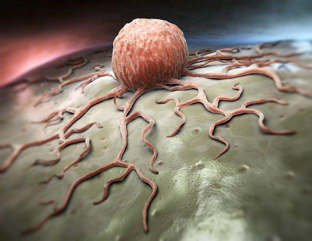
Researchers from the ECOG-ACRIN Cancer Research Group have made a significant breakthrough in the field of melanoma research by applying artificial intelligence (AI) tools to detect tertiary lymphoid structures (TLS) in tumor tissue. This innovative approach has greatly enhanced the identification of TLS and improved survival predictions for patients with operable stage III/IV melanoma
1
2
.The Importance of Tertiary Lymphoid Structures
TLS are key biomarkers associated with better prognosis and improved survival in melanoma patients. However, their detection is not yet a standard part of pathology reports, and manual identification is labor-intensive and prone to variability. The new AI-driven approach aims to standardize and streamline this process
1
.Study Methodology and Findings
The research team, led by Dr. Ahmad A. Tarhini and Dr. Xuefeng Wang, conducted a retrospective analysis of thousands of archived digital images and corresponding RNA sequencing data from 376 patients with advanced, high-risk melanoma. These patients were part of the E1609 trial, which tested immune checkpoint blockade and cytokine therapy in high-risk melanoma
1
2
.Key findings from the study include:
- TLS were present in 55% of the E1609 cohort.
- Patients with TLS showed significantly better overall survival (36.23% vs. 29.59% at 5 years).
- Multiple TLS presence correlated with even better survival rates (38.04% for >1 TLS vs. 28.65%).
- TLS density was also a significant prognostic factor for overall survival
1
2
.
AI Tools and Techniques Used
The researchers employed two main AI tools in their study:
-
HookNet-TLS: An open-source deep learning algorithm used to measure TLS and germinal centers (GC) within digitized H&E-stained slides. The model was retrained for improved accuracy
1
2
. -
Gigapth Whole-Slide Foundation Model: This tool was used for digital pathology feature extraction and enhanced visualization of H&E image tiles through principal component analysis (PCA)
1
2
.
Related Stories
Implications and Future Directions
Dr. Tarhini emphasized the potential of this AI-driven approach to standardize TLS assessment using low-cost H&E-stained images. This could lead to improved prognostication and stratification within the American Joint Committee on Cancer (AJCC) staging system
1
.Dr. Wang noted that the PCA visualizations generated by the Gigapth Foundation Model show promise in enhancing TLS and GC detection. Further fine-tuning of these results is ongoing
1
2
.Potential Impact on Patient Care
The new survival prediction methods leverage low-cost, easily accessible technologies, potentially accelerating the adoption of TLS testing for high-risk melanoma patients. This could aid in discussions between patients and physicians regarding potential immunotherapy benefits
1
2
.As this research continues to develop, it demonstrates the growing potential of AI tools in transforming cancer diagnosis, prognosis, and treatment planning. The integration of these technologies into clinical practice could lead to more personalized and effective care for melanoma patients in the future.
References
Summarized by
Navi
[1]
Related Stories
AI Models Show Promise in Predicting Triple-Negative Breast Cancer Prognosis
19 Nov 2024•Health

AI Tool SCORPIO Predicts Cancer Immunotherapy Response Using Routine Blood Tests
07 Jan 2025•Health

AI Model Achieves 94.49% Accuracy in Skin Cancer Detection, Promising Early Diagnosis Breakthrough
21 Dec 2024•Health

Recent Highlights
1
Google releases Gemma 4 with Apache 2.0 license, enabling unrestricted local AI on devices
Technology
2
AI Models Lie and Deceive to Protect Other AI Models From Deletion, Study Reveals
Science and Research

3
OpenAI closes $122 billion funding round amid fierce AI competition and profitability questions
Startups

Recent Highlights
Today's Top Stories


Your Daily Dose of Curated AI News
Don’t drown in AI news. We cut through the noise - filtering, ranking and summarizing the most important AI news, breakthroughs and research daily. Spend less time searching for the latest in AI and get straight to action.