Swiss Researchers Develop AI Model for Automated MRI Image Segmentation
3 Sources
3 Sources
[1]
Researchers develop AI model to automatically segment MRI images
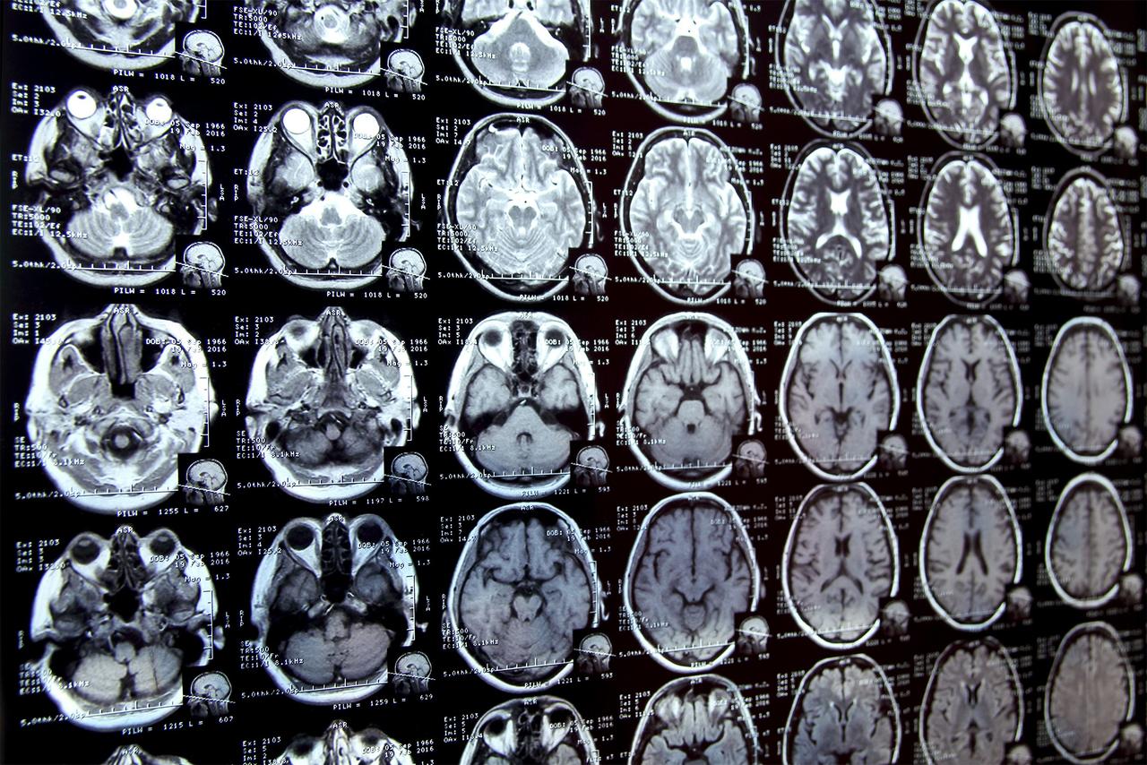
Research scientists in Switzerland have developed and tested a robust AI model that automatically segments major anatomic structures in MRI images, independent of sequence, according to a new study published today in Radiology, a journal of the Radiological Society of North America (RSNA). In the study, the model outperformed other publicly available tools. MRI provides detailed images of the human body and is essential for diagnosing various medical conditions, from neurological disorders to musculoskeletal injuries. For in-depth interpretation of MRI images, the organs, muscles and bones in the images are outlined or marked, which is known as segmenting. "MRI images have traditionally been manually segmented, which is a time-consuming process that requires intensive effort by radiologists and is subject to inter-reader variability," said Jakob Wasserthal, Ph.D., Radiology Department research scientist at University Hospital Basel in Basel, Switzerland. "Automated systems can potentially reduce radiologist's workload, minimize human errors and provide more consistent and reproducible results." Dr. Wasserthal and colleagues built an open-source automated segmentation tool called the TotalSegmentator MRI based on nnU-Net, a self-configuring framework that has set new standards in medical image segmentation. It adapts to any new dataset with minimal user intervention, automatically adjusting its architecture, preprocessing, and training strategies to optimize performance. A similar model for CT (TotalSegmentator CT) is being used by over 300,000 users worldwide to process over 100,000 CT images daily. In the retrospective study, the researchers trained TotalSegmentator MRI to provide sequence-independent segmentations of major anatomic structures using a randomly sampled dataset of 616 MRI and 527 CT exams. The training set included segmentations of 80 anatomic structures typically used for measuring volume, characterizing disease, surgical planning and opportunistic screening. "Our innovation was creating a large data set," Dr. Wasserthal said. "We used a lot more data and segmented many more organs, bones and muscles than has been previously done. Our model also works across different MRI scanners and image acquisition settings." To evaluate the model's performance, Dice scores -- which measure how similar two sets of data are -- were calculated between predicted segmentations and radiologist reference standards for segmentations. The model performed well across the 80 structures with a Dice score of 0.839 on an internal MRI test set. It also significantly outperformed two publicly available segmentation models (0.862 versus 0.838 and 0.560) and matched the performance of TotalSegmentator CT. "To our knowledge, our model is the only one that can automatically segment the highest number of structures on MRIs of any sequence," he said. "It's a tool that helps improve radiologists' work, makes measurements more precise and enables other measurements to be done that would have taken too much time to do manually." In addition to research and AI product development, Dr. Wasserthal said the model could potentially be used clinically for treatment planning, monitoring disease progression, and opportunistic screening.
[2]
AI model automatically segments MRI images, reducing radiologist workload
Research scientists in Switzerland have developed and tested a robust AI model that automatically segments major anatomic structures in MRI images, independent of sequence, according to a study published in Radiology. In the study, the model outperformed other publicly available tools. MRI provides detailed images of the human body and is essential for diagnosing various medical conditions, from neurological disorders to musculoskeletal injuries. For in-depth interpretation of MRI images, the organs, muscles and bones in the images are outlined or marked, which is known as segmenting. "MRI images have traditionally been manually segmented, which is a time-consuming process that requires intensive effort by radiologists and is subject to inter-reader variability," said Jakob Wasserthal, Ph.D., Radiology Department research scientist at University Hospital Basel in Basel, Switzerland. "Automated systems can potentially reduce a radiologist's workload, minimize human errors and provide more consistent and reproducible results." Dr. Wasserthal and colleagues built an open-source automated segmentation tool called the TotalSegmentator MRI based on nnU-Net, a self-configuring framework that has set new standards in medical image segmentation. It adapts to any new dataset with minimal user intervention, automatically adjusting its architecture, preprocessing, and training strategies to optimize performance. A similar model for CT (TotalSegmentator CT) is being used by over 300,000 users worldwide to process over 100,000 CT images daily. In the retrospective study, the researchers trained TotalSegmentator MRI to provide sequence-independent segmentations of major anatomic structures using a randomly sampled dataset of 616 MRI and 527 CT exams. The training set included segmentations of 80 anatomic structures typically used for measuring volume, characterizing disease, surgical planning and opportunistic screening. "Our innovation was creating a large data set," Dr. Wasserthal said. "We used a lot more data and segmented many more organs, bones and muscles than has been previously done. Our model also works across different MRI scanners and image acquisition settings." To evaluate the model's performance, Dice scores -- which measure how similar two sets of data are -- were calculated between predicted segmentations and radiologist reference standards for segmentations. The model performed well across the 80 structures with a Dice score of 0.839 on an internal MRI test set. It also significantly outperformed two publicly available segmentation models (0.862 versus 0.838 and 0.560) and matched the performance of TotalSegmentator CT. "To our knowledge, our model is the only one that can automatically segment the highest number of structures on MRIs of any sequence," he said. "It's a tool that helps improve radiologists' work, makes measurements more precise and enables other measurements to be done that would have taken too much time to do manually." In addition to research and AI product development, Dr. Wasserthal said the model could potentially be used clinically for treatment planning, monitoring disease progression, and opportunistic screening.
[3]
AI model automatically segments major structures in MRI images
Radiological Society of North AmericaFeb 18 2025 Research scientists in Switzerland have developed and tested a robust AI model that automatically segments major anatomic structures in MRI images, independent of sequence, according to a new study published today in Radiology, a journal of the Radiological Society of North America (RSNA). In the study, the model outperformed other publicly available tools. MRI provides detailed images of the human body and is essential for diagnosing various medical conditions, from neurological disorders to musculoskeletal injuries. For in-depth interpretation of MRI images, the organs, muscles and bones in the images are outlined or marked, which is known as segmenting. MRI images have traditionally been manually segmented, which is a time-consuming process that requires intensive effort by radiologists and is subject to inter-reader variability. Automated systems can potentially reduce radiologist's workload, minimize human errors and provide more consistent and reproducible results." Jakob Wasserthal, Ph.D., Radiology Department research scientist at University Hospital Basel in Basel, Switzerland Dr. Wasserthal and colleagues built an open-source automated segmentation tool called the TotalSegmentator MRI based on nnU-Net, a self-configuring framework that has set new standards in medical image segmentation. It adapts to any new dataset with minimal user intervention, automatically adjusting its architecture, preprocessing, and training strategies to optimize performance. A similar model for CT (TotalSegmentator CT) is being used by over 300,000 users worldwide to process over 100,000 CT images daily. In the retrospective study, the researchers trained TotalSegmentator MRI to provide sequence-independent segmentations of major anatomic structures using a randomly sampled dataset of 616 MRI and 527 CT exams. The training set included segmentations of 80 anatomic structures typically used for measuring volume, characterizing disease, surgical planning and opportunistic screening. "Our innovation was creating a large data set," Dr. Wasserthal said. "We used a lot more data and segmented many more organs, bones and muscles than has been previously done. Our model also works across different MRI scanners and image acquisition settings." To evaluate the model's performance, Dice scores-which measure how similar two sets of data are-were calculated between predicted segmentations and radiologist reference standards for segmentations. The model performed well across the 80 structures with a Dice score of 0.839 on an internal MRI test set. It also significantly outperformed two publicly available segmentation models (0.862 versus 0.838 and 0.560) and matched the performance of TotalSegmentator CT. "To our knowledge, our model is the only one that can automatically segment the highest number of structures on MRIs of any sequence," he said. "It's a tool that helps improve radiologists' work, makes measurements more precise and enables other measurements to be done that would have taken too much time to do manually." In addition to research and AI product development, Dr. Wasserthal said the model could potentially be used clinically for treatment planning, monitoring disease progression, and opportunistic screening. Radiological Society of North America Journal reference: Akinci D'Antonoli, T., et al. (2025). TotalSegmentator MRI: Robust Sequence-independent Segmentation of Multiple Anatomic Structures in MRI. Radiology. doi.org/10.1148/radiol.241613.
Share
Share
Copy Link
A new AI model called TotalSegmentator MRI, developed by Swiss researchers, can automatically segment major anatomic structures in MRI images across different sequences, potentially reducing radiologists' workload and improving diagnostic accuracy.

Swiss Researchers Develop Advanced AI Model for MRI Image Segmentation
Researchers at the University Hospital Basel in Switzerland have made a significant breakthrough in medical imaging technology with the development of a new AI model called TotalSegmentator MRI. This innovative tool automatically segments major anatomic structures in MRI images, regardless of the sequence used, potentially revolutionizing the field of radiology
1
.The Challenge of MRI Image Segmentation
MRI (Magnetic Resonance Imaging) is a crucial diagnostic tool in modern medicine, providing detailed images of the human body for various medical conditions. However, the process of segmenting these images - outlining organs, muscles, and bones - has traditionally been a manual, time-consuming task prone to human error and inter-reader variability
2
.TotalSegmentator MRI: A Game-Changing Solution
Dr. Jakob Wasserthal and his team at the University Hospital Basel have addressed this challenge by developing TotalSegmentator MRI, an open-source automated segmentation tool. Built on the nnU-Net framework, this AI model can adapt to new datasets with minimal user intervention, automatically optimizing its performance
3
.Key Features and Innovations
- Large-scale dataset: The model was trained on a diverse dataset of 616 MRI and 527 CT exams, encompassing 80 anatomic structures.
- Sequence independence: TotalSegmentator MRI works across different MRI scanners and image acquisition settings.
- Comprehensive segmentation: The model can segment the highest number of structures on MRIs of any sequence.
Related Stories


Performance and Validation
The researchers evaluated the model's performance using Dice scores, which measure the similarity between predicted segmentations and radiologist reference standards. TotalSegmentator MRI achieved impressive results:
- Dice score of 0.839 on an internal MRI test set
- Outperformed two publicly available segmentation models (0.862 vs 0.838 and 0.560)
- Matched the performance of its CT counterpart, TotalSegmentator CT
Potential Applications and Impact
The development of TotalSegmentator MRI has far-reaching implications for both research and clinical practice:
- Reduced radiologist workload: Automated segmentation can significantly decrease the time and effort required for image analysis.
- Improved consistency: The AI model minimizes human errors and provides more reproducible results.
- Enhanced measurements: The tool enables more precise measurements and facilitates analyses that were previously too time-consuming to perform manually.
- Clinical applications: Potential uses include treatment planning, monitoring disease progression, and opportunistic screening.
As the field of AI in medical imaging continues to advance, tools like TotalSegmentator MRI are poised to play a crucial role in improving diagnostic accuracy, streamlining workflows, and ultimately enhancing patient care.
References
Summarized by
Navi
[1]
[3]
Related Stories
Recent Highlights
1
OpenAI shuts down Sora video app after six months, ending Disney's $1 billion investment deal
Technology

2
AI-Generated Val Kilmer to Posthumously Appear in As Deep as the Grave After His Death
Entertainment and Society

3
Supermicro Co-Founder Indicted in $2.5 Billion Nvidia AI Chip Smuggling Scheme to China
Policy and Regulation

Recent Highlights
Today's Top Stories


Your Daily Dose of Curated AI News
Don’t drown in AI news. We cut through the noise - filtering, ranking and summarizing the most important AI news, breakthroughs and research daily. Spend less time searching for the latest in AI and get straight to action.