2 Sources
[1]
New AI spots hypertrophic cardiomyopathy obstruction from standard ultrasounds
Mayo Clinic researchers have developed and externally validated an artificial intelligence (AI) model that can identify a potentially significant heart obstruction from routine ultrasound videos without relying on specialized Doppler imaging. The technology could help clinicians flag patients with hypertrophic cardiomyopathy (HCM) who may need additional testing, particularly in settings where specialized echocardiography expertise is limited. Study findings are published in Circulation: Cardiovascular Imaging. HCM is a genetic condition that causes the heart muscle to become abnormally thick. About two-thirds of these patients develop left ventricular outflow tract (LVOT) obstruction, which restricts blood leaving the heart, causing symptoms such as chest pain and shortness of breath with exertion or when lying flat. Knowing which patients develop LVOT obstruction is important because it influences treatment decisions and long-term management for patients with HCM. "Measuring LVOT obstruction typically requires Doppler echocardiography, which depends on precise ultrasound-beam alignment and operator expertise," says Imon Banerjee, Ph.D., an AI researcher at Mayo Clinic in Phoenix and senior author of the study. "We wanted to determine whether AI could recognize subtle patterns that are imperceptible to the human eye in routinely acquired B-mode ultrasound videos and identify patients with LVOT obstruction earlier, enabling timely confirmatory Doppler evaluation and referral when appropriate." The study included 1,833 patients in the Mayo Clinic cohort. The model was tested in 275 patients and externally validated in 46 patients from a hospital in South Korea. The AI model used only resting, non-Doppler ultrasound videos to predict whether a patient had a potentially significant obstruction to blood leaving the heart. Researchers found that combining information from three standard ultrasound views improved the model's ability to distinguish patients with elevated LVOT gradients. The model also helped identify obstruction that may only appear when the heart is under stress. The model maintained strong performance in the South Korean group despite substantial differences between that population and the patients used to develop the model, supporting further study of the technology across different patient populations and clinical settings. In a subset of cases, the AI model identified obstruction more accurately than two expert echocardiographers who reviewed the same non-Doppler images. The findings highlight how difficult it can be to recognize LVOT obstruction from routine two-dimensional images without Doppler measurements. "This technology is intended to complement, not replace, Doppler echocardiography," Dr. Banerjee says. "By enabling earlier identification of patients with potential LVOT obstruction, it could escalate timely detection and prompt confirmatory Doppler measurements, stress testing, or referral to an HCM specialty center. It also could support evaluation using portable ultrasound or in settings where comprehensive Doppler assessment may not be readily available, helping expand access to earlier screening and risk assessment." Dr. Banerjee notes that the next steps include additional prospective validation across broader clinical settings, ultrasound platforms and patient populations.
[2]
Mayo Clinic AI model helps clinicians detect heart obstruction using routine ultrasound images | Newswise
PHOENIX -- Mayo Clinic researchers have developed and externally validated an artificial intelligence (AI) model that can identify a potentially significant heart obstruction from routine ultrasound videos without relying on specialized Doppler imaging. The technology could help clinicians flag patients with hypertrophic cardiomyopathy (HCM) who may need additional testing, particularly in settings where specialized echocardiography expertise is limited. Study findings are published in Circulation: Cardiovascular Imaging. HCM is a genetic condition that causes the heart muscle to become abnormally thick. About two-thirds of these patients develop left ventricular outflow tract (LVOT) obstruction, which restricts blood leaving the heart, causing symptoms such as chest pain and shortness of breath with exertion or when lying flat. Knowing which patients develop LVOT obstruction is important because it influences treatment decisions and long-term management for patients with HCM. "Measuring LVOT obstruction typically requires Doppler echocardiography, which depends on precise ultrasound-beam alignment and operator expertise," says Imon Banerjee, Ph.D., an AI researcher at Mayo Clinic in Phoenix and senior author of the study. "We wanted to determine whether AI could recognize subtle patterns that are imperceptible to the human eye in routinely acquired B-mode ultrasound videos and identify patients with LVOT obstruction earlier, enabling timely confirmatory Doppler evaluation and referral when appropriate." The study included 1,833 patients in the Mayo Clinic cohort. The model was tested in 275 patients and externally validated in 46 patients from a hospital in South Korea. The AI model used only resting, non-Doppler ultrasound videos to predict whether a patient had a potentially significant obstruction to blood leaving the heart. Researchers found that combining information from three standard ultrasound views improved the model's ability to distinguish patients with elevated LVOT gradients. The model also helped identify obstruction that may only appear when the heart is under stress. The model maintained strong performance in the South Korean group despite substantial differences between that population and the patients used to develop the model, supporting further study of the technology across different patient populations and clinical settings. In a subset of cases, the AI model identified obstruction more accurately than two expert echocardiographers who reviewed the same non-Doppler images. The findings highlight how difficult it can be to recognize LVOT obstruction from routine two-dimensional images without Doppler measurements. "This technology is intended to complement, not replace, Doppler echocardiography," Dr. Banerjee says. "By enabling earlier identification of patients with potential LVOT obstruction, it could escalate timely detection and prompt confirmatory Doppler measurements, stress testing, or referral to an HCM specialty center. It also could support evaluation using portable ultrasound or in settings where comprehensive Doppler assessment may not be readily available, helping expand access to earlier screening and risk assessment." Dr. Banerjee notes that the next steps include additional prospective validation across broader clinical settings, ultrasound platforms and patient populations. The list of authors and disclosures may be found in the article, Beyond Doppler: Scalable AI Detection of LVOT Obstruction in HCM. This study received no external funding. ### About Mayo Clinic Mayo Clinic is a nonprofit organization committed to innovation in clinical practice, education and research, and providing compassion, expertise and answers to everyone who needs healing. Visit the Mayo Clinic News Network for additional Mayo Clinic news.
Share
Copy Link
Mayo Clinic developed an AI model that identifies left ventricular outflow tract obstruction in hypertrophic cardiomyopathy patients using standard ultrasound videos without specialized Doppler imaging. Tested on 1,833 patients and validated in South Korea, the model outperformed expert echocardiographers in detecting obstructions, potentially enabling earlier diagnosis in resource-limited settings.
Mayo Clinic researchers developed an AI model that detects left ventricular outflow tract obstruction in hypertrophic cardiomyopathy patients using only routine ultrasound videos, eliminating the need for specialized Doppler echocardiography
1
. Published in Circulation: Cardiovascular Imaging, this technology addresses a critical gap in cardiac diagnostics by identifying subtle patterns imperceptible to the human eye in B-mode ultrasound images2
. The model trained on 1,833 patients in the Mayo Clinic cohort, tested on 275 patients, and externally validated in 46 patients from a South Korean hospital, demonstrating robust performance across different populations1
.Hypertrophic cardiomyopathy affects approximately two-thirds of patients who develop left ventricular outflow tract (LVOT) obstruction, restricting blood flow from the heart and causing chest pain and shortness of breath
2
. Traditional diagnosis requires Doppler echocardiography, which depends on precise ultrasound-beam alignment and operator expertise, creating barriers in resource-limited settings where specialized echocardiography expertise may not be readily available1
. Senior author Imon Banerjee, Ph.D., an AI researcher at Mayo Clinic in Phoenix, explained the team wanted to determine whether AI-assisted cardiac diagnostics could recognize patterns in routinely acquired ultrasound videos to enable earlier diagnosis and timely confirmatory Doppler evaluation2
.
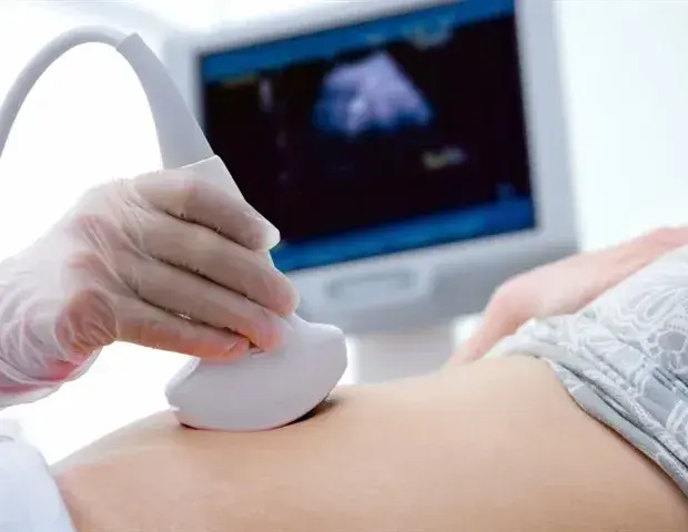
Source: News-Medical
The AI model achieved notable results by combining information from three standard ultrasound views, improving its ability to distinguish patients with elevated LVOT gradients
1
. In a subset of cases, the model identified obstruction more accurately than two expert echocardiographers who reviewed the same non-Doppler images, highlighting how difficult recognizing LVOT obstruction can be from routine two-dimensional images without Doppler measurements2
. The technology also helped identify obstruction that may only appear when the heart is under stress, expanding diagnostic capabilities beyond resting conditions1
.Related Stories
Dr. Banerjee emphasized the technology is intended to complement, not replace, Doppler echocardiography, enabling earlier identification of patients with potential LVOT obstruction and prompting confirmatory testing, stress testing, or referral to an HCM specialty center
2
. The model maintained strong performance in the external validation despite substantial differences between the South Korean population and the development cohort, supporting broader clinical implementation across different patient populations and settings1
. This capability could expand access to earlier screening and risk assessment using portable ultrasound in environments where comprehensive Doppler assessment is not readily available, potentially transforming how clinicians approach hypertrophic cardiomyopathy detection2
. Next steps include additional prospective validation across broader clinical settings, ultrasound platforms, and patient populations to establish the model's utility in real-world practice1
.Summarized by
Navi
17 Jul 2025•Health

10 Jul 2025•Health

23 Apr 2025•Health

1
Technology

2
Technology

3
Technology