AI analyzes retinal photos to predict Alzheimer's disease risk decades before symptoms appear
2 Sources
[1]
Retinal Photos and AI Predict Early Alzheimer's Risk
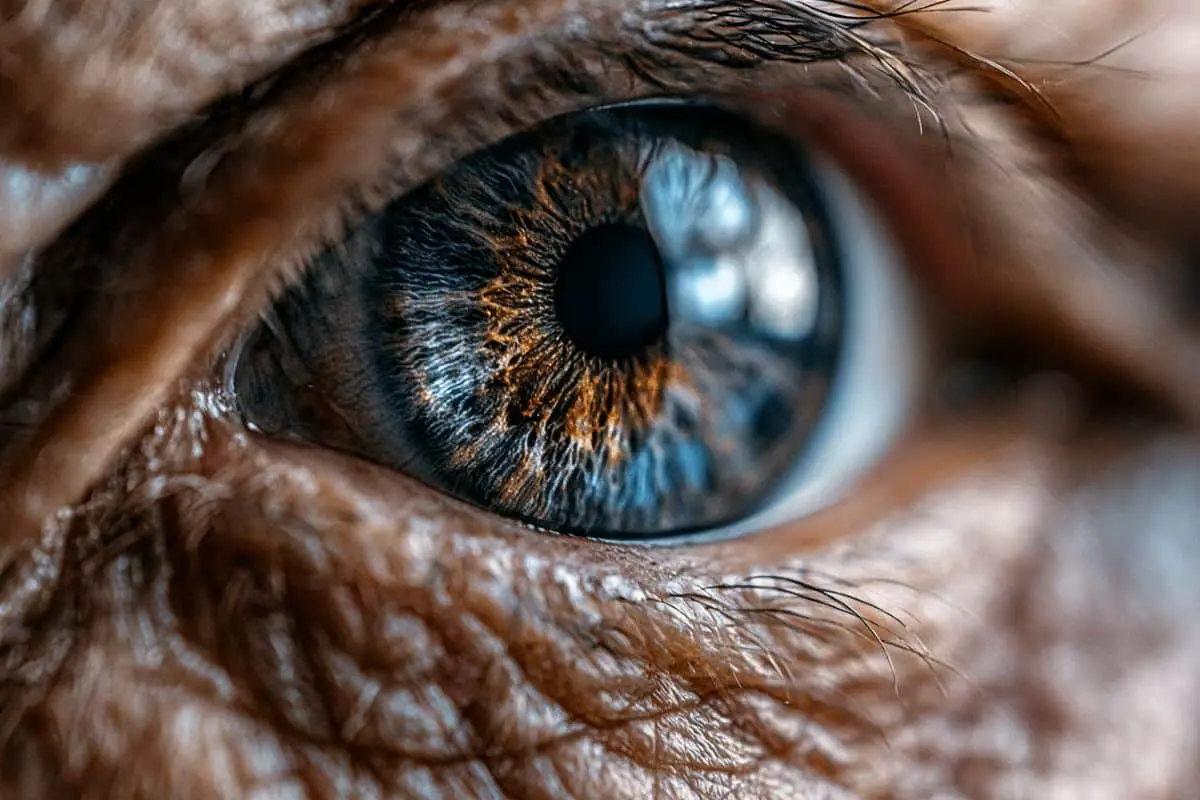
Summary: A new study that used artificial intelligence to analyze routine eye photographs has unlocked a cheap, non-invasive method for predicting major Alzheimer's disease risk factors decades before clinical symptoms appear. By training machine learning models on retinal photographs from over 40,000 patients in a UK-based databank, the research team successfully mapped specific regions of the eye, such as the retinal arteries and optic nerve, to biological and lifestyle risk factors associated with Alzheimer's vulnerability. The AI accurately predicted characteristics including biological sex, blood pressure, smoking, alcohol consumption, and insomnia. Because the retina serves as a direct extension of the central nervous system, these common, low-cost images act as an "integrated biological sensor" of a patient's cumulative neurovascular damage, opening a vital window for early lifestyle and medical interventions before irreversible brain damage occurs. Key Facts * The Ocular Window: Retinal morphology (specifically arteries and the optic nerve) provides measurable indicators of neurovascular integrity, acting as an organic sensor for Alzheimer's vulnerability. * Massive Databank Trial: The machine learning model was trained and validated using retinal images from more than 40,000 patients archived in a major United Kingdom database. * Objective Risk Mapping: The AI successfully bypasses unreliable patient self-reports to objectively identify lifestyle and biological risks, including high blood pressure, smoking, alcohol use, and insomnia. * Decade-Early Intervention: Because Alzheimer's pathologies develop over decades, this low-cost screening identifies at-risk patients long before late-stage, irreversible brain damage occurs. * Ubiquitous & Cost-Effective: Unlike cost-prohibitive MRIs or PET scans, retinal photography is already widely performed during routine eye exams, diabetes screenings, and glaucoma checks. Source: University of Florida Often called "the window to the soul," the eyes may also offer clues about something less poetic but just as important: the health of the brain. A new study of tens of thousands of patients revealed that cheap, simple and common photographs of the retina at the back of the eye can accurately predict many of the most common risk factors that are associated with developing Alzheimer's disease. "We know that Alzheimer's disease develops over decades, but most of the diagnostic tools focus on late stage pathology when it is too late to intervene," said Ruogu Fang, Ph.D., a professor of biomedical engineering at the University of Florida who led the new study. "By looking at novel biomarkers, like retinal health, we offer new opportunities to identify patients at risk, offer appropriate tests and encourage them to develop healthy lifestyles to mitigate their risk." Fang and her collaborators, including UF's Adam Woods, Ph.D., and Meta researcher Yunchao Yang, Ph.D., published their findings June 16 in the Journal of Alzheimer's Disease. Many patients routinely have pictures of their eyes taken. Those with diabetes, glaucoma or cataracts will have many retinal photographs taken over the years. Even regular eye exams for prescription glasses can capture photos. That near-ubiquity makes analyzing retinal photographs simple and low-cost compared to other, more expensive technologies like MRIs. By using machine learning to analyze these retinal photographs from more than 40,000 patients in a United Kingdom-based patient databank, Fang's group was able to identify regions of the retina associated with Alzheimer's risk factors, such as the arteries and optical nerve. "With the assistance of AI, we are now able to identify subtle retinal variations that were formerly overlooked across thousands of subjects, which may function as reliable indicators of future disease risk," said Seowung Leem, a doctoral student at UF and first author of the publication. The AI model accurately predicted biological characteristics like sex or blood pressure as well as lifestyle factors associated with developing Alzheimer's, such as smoking, alcohol use and even insomnia. While many of these factors are captured in patients' medical charts, those records are often incomplete. Some, like alcohol and smoking, rely on unreliable self-reports. So retinal photographs may provide another, more objective way to detect these risk factors. Plus, the retinal images can capture damage accumulated over the years, which will vary between patients who share similar risk factors. "Retinal morphology could provide measurable indicators of neurovascular integrity, which is highly relevant to Alzheimer's disease vulnerability," said Fang, who is also affiliated with the McKnight Brain Institute. "In this sense, retinal imaging functions less as a surrogate questionnaire and more as an integrated biological sensor of cumulative risk." Fang's group has already established that retinal photographs can detect active cases of Alzheimer's disease. But scientists now believe the disease progresses over many years, even decades. So identifying early risk factors could better identify patients who could respond to earlier interventions -- including protective lifestyle changes, some medications or even brain training -- before irreversible damage to the brain takes place. Funding: The work was supported in part by the National Science Foundation. Key Questions Answered: Editorial Notes: * This article was edited by a Neuroscience News editor. * Journal paper reviewed in full. * Additional context added by our staff. About this AI and Alzheimer's disease research news Author: Eric Hamilton Source: University of Florida Contact: Eric Hamilton - University of Florida Image: The image is credited to Neuroscience News Original Research: Closed access. "Prediction of Alzheimer's disease risk factors from retinal images via deep learning: Development and validation of biologically relevant morphological associations in the UK Biobank" by Ruogu Fang et al. Journal of Alzheimer's Disease DOI:10.1177/13872877261457650 Abstract Prediction of Alzheimer's disease risk factors from retinal images via deep learning: Development and validation of biologically relevant morphological associations in the UK Biobank Background The systemic, metabolic, lifestyle factors have established associations with Alzheimer's disease (AD) through epidemiologic and AD-specific biomarker studies. Whether colored fundus photography (CFP) contains retinal structural signatures corresponding to these AD-related risk domains remains unclear. Objective To determine whether deep learning (DL) models can predict 12 AD-related risk factors from CFP and to characterize the retinal structures underlying these predictions, thereby assessing whether CFP reflects pathways to AD vulnerability. Methods Using 62,876 CFPs from 44,501 unique participants from the UK Biobank, DL models were trained to predict 12 factors linked to AD pathology or incidence: 6 categorical (sex, smoking, sleeplessness, economic status, alcohol use, depression) and 6 continuous (age, age at completing education, body mass index, systolic, diastolic blood pressure, HbA1c). Model performance, model saliency, and saliency-derived scores (CAM-Score) were evaluated and compared to retinal morphometry. The scores were also compared between incident-AD cases (average 8.55 years before onset) and matched controls. Results Predictive performance of DL ranged from AUROC between 0.5654 and 0.9480 for categorical factors and R between -0.0291 and 0.7620 for continuous factors, outperforming most of the morphometry-based machine learning models. Saliency-based score consistently highlighted biologically meaningful regions, particularly the optic nerve head and retinal vasculature. It also aligned with present morphometric variations. Several saliency-based scores differed significantly between incident AD and matched controls, suggesting potential overlap between retinal correlates of AD-related risk factors and preclinical AD-associated changes. Conclusions CFP encodes retinal signatures linked to AD risk factors. Although not diagnostic, DL-derived retinal representations may uncover biologically meaningful risk-related structural changes mirroring the potential AD vulnerability.
[2]
Retinal photographs may reveal early Alzheimer's disease risk
University of FloridaJun 16 2026 Often called "the window to the soul," the eyes may also offer clues about something less poetic but just as important: the health of the brain. A new study of tens of thousands of patients revealed that cheap, simple and common photographs of the retina at the back of the eye can accurately predict many of the most common risk factors that are associated with developing Alzheimer's disease. "We know that Alzheimer's disease develops over decades, but most of the diagnostic tools focus on late stage pathology when it is too late to intervene," said Ruogu Fang, Ph.D., a professor of biomedical engineering at the University of Florida who led the new study. "By looking at novel biomarkers, like retinal health, we offer new opportunities to identify patients at risk, offer appropriate tests and encourage them to develop healthy lifestyles to mitigate their risk." Fang and her collaborators, including UF's Adam Woods, Ph.D., and Meta researcher Yunchao Yang, Ph.D., published their findings June 16 in the Journal of Alzheimer's Disease. The work was supported in part by the National Science Foundation. Many patients routinely have pictures of their eyes taken. Those with diabetes, glaucoma or cataracts will have many retinal photographs taken over the years. Even regular eye exams for prescription glasses can capture photos. That near-ubiquity makes analyzing retinal photographs simple and low-cost compared to other, more expensive technologies like MRIs. By using machine learning to analyze these retinal photographs from more than 40,000 patients in a United Kingdom-based patient databank, Fang's group was able to identify regions of the retina associated with Alzheimer's risk factors, such as the arteries and optical nerve. "With the assistance of AI, we are now able to identify subtle retinal variations that were formerly overlooked across thousands of subjects, which may function as reliable indicators of future disease risk," said Seowung Leem, a doctoral student at UF and first author of the publication. The AI model accurately predicted biological characteristics like sex or blood pressure as well as lifestyle factors associated with developing Alzheimer's, such as smoking, alcohol use and even insomnia. While many of these factors are captured in patients' medical charts, those records are often incomplete. Some, like alcohol and smoking, rely on unreliable self-reports. So retinal photographs may provide another, more objective way to detect these risk factors. Plus, the retinal images can capture damage accumulated over the years, which will vary between patients who share similar risk factors. "Retinal morphology could provide measurable indicators of neurovascular integrity, which is highly relevant to Alzheimer's disease vulnerability," said Fang, who is also affiliated with the McKnight Brain Institute. "In this sense, retinal imaging functions less as a surrogate questionnaire and more as an integrated biological sensor of cumulative risk." Fang's group has already established that retinal photographs can detect active cases of Alzheimer's disease. But scientists now believe the disease progresses over many years, even decades. So identifying early risk factors could better identify patients who could respond to earlier interventions - including protective lifestyle changes, some medications or even brain training - before irreversible damage to the brain takes place.
Share
Copy Link
University of Florida researchers trained machine learning models on retinal photographs from over 40,000 patients to identify early Alzheimer's disease risk factors. The AI-powered analysis detected biological and lifestyle indicators like blood pressure, smoking, and insomnia by examining the retina's arteries and optic nerve, offering a low-cost alternative to expensive brain scans.
AI Transforms Routine Eye Photos Into Alzheimer's Risk Predictors
Researchers at the University of Florida have developed an AI model that analyzes retinal photographs to predict Alzheimer's disease risk factors decades before clinical symptoms emerge. By training machine learning algorithms on retinal images from more than 40,000 patients in a United Kingdom-based databank, the team successfully identified specific eye regions—particularly the retinal arteries and optic nerve—that correlate with biological and lifestyle vulnerabilities associated with Alzheimer's development
1
. The findings, published June 16 in the Journal of Alzheimer's Disease, demonstrate how AI-powered analysis of retinal photographs can serve as a low-cost non-invasive tool for early detection and intervention2
.
Source: Neuroscience News
"We know that Alzheimer's disease develops over decades, but most of the diagnostic tools focus on late stage pathology when it is too late to intervene," said Ruogu Fang, Ph.D., a professor of biomedical engineering at the University of Florida who led the study. "By looking at novel biomarkers, like retinal health, we offer new opportunities to identify patients at risk, offer appropriate tests and encourage them to develop healthy lifestyles to mitigate their risk"
1
.Machine Learning Detects Subtle Retinal Variations Linked to Risk
The AI model accurately predicted biological characteristics including sex and blood pressure, as well as lifestyle factors such as smoking, alcohol use, and insomnia—all known contributors to early Alzheimer's disease risk
2
. "With the assistance of AI, we are now able to identify subtle retinal variations that were formerly overlooked across thousands of subjects, which may function as reliable indicators of future disease risk," explained Seowung Leem, a doctoral student at the University of Florida and first author of the publication1
.Because the retina serves as a direct extension of the central nervous system, retinal photographs function as an "integrated biological sensor" that captures cumulative neurovascular damage over time. This approach bypasses unreliable patient self-reports often found in medical charts, providing objective measurements that vary between individuals even when they share similar risk profiles
2
.Cost-Effective Alternative to Expensive Brain Imaging
Unlike cost-prohibitive MRIs or PET scans, retinal photographs are already captured routinely during eye exams for prescription glasses, diabetes screenings, and glaucoma checks. This near-ubiquity makes the approach far more accessible for widespread screening
1
. Patients with diabetes, glaucoma, or cataracts typically have multiple retinal photographs taken over years, creating extensive archives that machine learning models can analyze to predict Alzheimer's risk factors2
."Retinal morphology could provide measurable indicators of neurovascular integrity, which is highly relevant to Alzheimer's disease vulnerability," Fang noted. "In this sense, retinal imaging functions less as a surrogate questionnaire and more as an integrated biological sensor of cumulative risk"
1
.
Source: News-Medical
Related Stories


Opening Windows for Preventive Interventions
The work, supported in part by the National Science Foundation and conducted in collaboration with UF's Adam Woods, Ph.D., and Meta researcher Yunchao Yang, Ph.D., builds on Fang's previous research showing that retinal biomarkers can detect active Alzheimer's cases
2
. Because Alzheimer's pathologies develop over many years, identifying at-risk patients early could enable protective lifestyle changes, medications, or brain training before irreversible damage occurs. This positions AI analysis of routine eye images as a practical screening method that could identify millions of vulnerable individuals who might benefit from preventive strategies, potentially altering the trajectory of cognitive decline before symptoms manifest.References
Summarized by
Navi
[1]
[2]
Related Stories
Recent Highlights
1
UN Warns AI Development Outpaces Governance as Global Dialogue Opens in Geneva
Policy and Regulation

2
OpenAI proposes giving US government 5% stake worth $42.6 billion amid regulatory pressure
Policy and Regulation

3
CIA Director calls cutting-edge AI 'digital nuclear weapons' as US tightens export controls
Policy and Regulation

Recent Highlights
Today's Top Stories


Don’t drown in AI news. We cut through the noise - filtering, ranking and summarizing the most important AI news, breakthroughs and research daily. Follow topics that matter to you and stay ahead.


