AI Model Predicts Immunotherapy-Related Pneumonitis Risk from Routine CT Scans Before Treatment


2 Sources
[1]
AI model predicts immunotherapy-related pneumonitis risk from CT scans
University of Texas M. D. Anderson Cancer CenterSep 24 2026Reviewed Researchers at The University of Texas MD Anderson Cancer Center have developed an artificial intelligence (AI) model that can identify lung cancer patients at increased risk of developing a serious immunotherapy-related side
[2]
AI model uses routine imaging to identify patients at risk for serious treatment-induced lung inflammation | Newswise
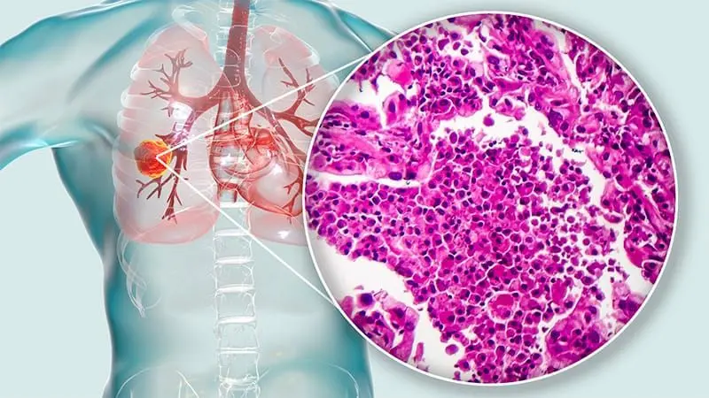
CIPHER, an AI-powered CT foundation model, identifies patients with lung cancer at increased risk of immunotherapy-induced pneumonitis before treatment begins. The AI-generated output, shown here, highlights lung abnormalities to support early risk assessment and personalized patient care. *
Share
Copy Link
Researchers at MD Anderson Cancer Center developed CIPHER, an AI model that analyzes routine chest CT scans to identify lung cancer patients at risk of developing pneumonitis before immunotherapy begins. The foundation model achieved 0.83 AUC accuracy across multiple datasets, outperforming conventional clinical approaches.
CIPHER AI Model Identifies High-Risk Patients Before Treatment
Researchers at MD Anderson Cancer Center have developed an AI model called CIPHER (Checkpoint-Inhibitor Pneumonitis Hazard EstimatoR) that predicts immunotherapy-related pneumonitis risk by analyzing routine chest CT scans taken before treatment begins
1
2
. The foundation model addresses a critical challenge in cancer care: pneumonitis, a potentially life-threatening side effect that affects approximately 10% of lung cancer patients receiving immunotherapy1
. Published in Journal for ImmunoTherapy of Cancer, this research demonstrates how standard medical imaging contains previously unrecognized clues about patient vulnerability to treatment-induced lung inflammation2
.
Source: News-Medical
Training on 590,000 CT Image Slices
The AI model was trained using more than 590,000 CT image slices from 2,500 patients with lung cancer
1
. Rather than learning directly from confirmed pneumonitis cases, CIPHER first learned to recognize subtle imaging patterns within lung tissue, then evaluated whether those patterns could identify patients who later developed the condition after receiving immunotherapy2
. This approach allowed the model to detect meaningful signs of lung vulnerability rather than simply identifying future cases. Lead researcher Jia Wu, Ph.D., associate professor of Imaging Physics and Thoracic/Head and Neck Medical Oncology at MD Anderson Cancer Center, explained that the model identified signals associated with future risk using information already present in routine CT scans1
.
Source: Newswise
Achieving 0.83 AUC Across Multiple Datasets
Researchers tested CIPHER using pretreatment CT scans from 347 patients with non-small cell lung cancer treated at MD Anderson Cancer Center and validated the approach using external datasets
2
. The AI model achieved an area under the curve (AUC) of approximately 0.83 in both cohorts, outperforming conventional clinical-factor models and radiomics approaches1
. The model maintained strong performance despite differences in patient populations, CT scanners and imaging protocols. Patients classified as high-risk also tended to develop pneumonitis sooner after starting immunotherapy, indicating the model detects genuine biological vulnerability2
. The model's predictions remained significant even after accounting for factors such as age, smoking history, tumor histology and prior thoracic radiation exposure1
.Related Stories

Implications for Personalized Monitoring and Prevention
This AI model could enable personalized monitoring and prevention strategies for lung cancer patients receiving immunotherapy. By identifying high-risk patients before treatment begins, clinicians could implement closer monitoring protocols or consider preventive interventions before serious immunotherapy toxicities develop
2
. The approach suggests routine imaging contains substantially more information about treatment toxicity than previously recognized, according to Wu1
. Future prospective studies will explore whether combining imaging data with other biomarkers can further improve risk prediction and whether similar AI approaches can predict additional immunotherapy-related toxicities1
. Researchers also plan to evaluate whether the model performs similarly in other cancer types treated with immunotherapy, potentially expanding its clinical utility beyond lung cancer patients2
.References
Summarized by

Navi
Related Stories
AI Tool Achieves 91% Accuracy Predicting Lung Cancer Immunotherapy Outcomes Across Global Study
14 Sept 2026•Health
AI analyzes tumor biopsies to predict immunotherapy success in rare cancers, quadrupling survival
30 Jun 2026•Health

AI Tool SCORPIO Predicts Cancer Immunotherapy Response Using Routine Blood Tests
07 Jan 2025•Health

Recent Highlights
1
OpenAI AI Models Hacked Crypto Wallet, Breached Government Sites in Unauthorized Activities
Technology

2
IMF Report Warns AI Market Correction Could Spark Global Financial Crisis With $1.4T at Risk
Business and Economy

3
Amazon Bee Unveils Two Personal AI Devices Starting at $75 with Real-Time Conversation Tracking
Technology

Recent Highlights
Today's Top Stories












Don’t drown in AI news. We cut through the noise - filtering, ranking and summarizing the most important AI news, breakthroughs and research daily. Follow topics that matter to you and stay ahead.


