Revolutionary Machine Learning Method Enhances MRI Imaging Efficiency


3 Sources
[1]
Better MRI videos thanks to new machine learning method
Using smartly trained neural networks, researchers at TU Graz have succeeded in generating precise real-time images of the beating heart from just a few MRI measurement data. Other MRI applications can also be accelerated using this procedure. Medical imaging using magnetic resonance imaging (MRI)
[2]
Better MRI videos thanks to new machine learning method
Using smartly trained neural networks, researchers at TU Graz have succeeded in generating precise real-time images of the beating heart from just a few MRI measurement data. Other MRI applications can also be accelerated using this procedure. The study is published in Magnetic Resonance in
[3]
Innovative machine learning technique transforms MRI imaging efficiency
TU GrazSep 30 2024 Using smartly trained neural networks, researchers at TU Graz have succeeded in generating precise real-time images of the beating heart from just a few MRI measurement data. Other MRI applications can also be accelerated using this procedure. Medical imaging using magnetic
Share
Copy Link
Researchers at TU Graz have developed a novel machine learning technique that generates precise real-time MRI images of the beating heart using minimal data, potentially revolutionizing medical imaging practices.

Breakthrough in MRI Technology
Researchers at Graz University of Technology (TU Graz) have made a significant advancement in magnetic resonance imaging (MRI) technology. Led by Martin Uecker and Moritz Blumenthal from the Institute of Biomedical Imaging, the team has developed a machine learning method that generates precise real-time images of the beating heart using minimal MRI measurement data
1
.Innovative Self-Supervised Learning Approach
The novel technique employs self-supervised learning methods to train neural networks for MRI imaging. Unlike traditional approaches that require perfect training images, this method uses a subset of the initial MRI data to reconstruct images
2
.Moritz Blumenthal explains the process: "We divided the measurement data provided by the MRI device into two portions. From the first, larger data portion, our machine learning model reconstructs the image. It then attempts to calculate the second portion of the measurement data withheld from it on the basis of the image"
3
.Iterative Improvement and Consistency
The system undergoes multiple rounds of reconstruction and validation. If the model fails to accurately calculate the withheld data, it updates itself and creates an improved image variant. This iterative process continues until a consistent result is achieved, allowing the system to learn what constitutes a good MRI image
1
.Related Stories

Potential Applications and Benefits
While the process is ready for application, practical implementation may take some time. The method has potential applications beyond cardiac imaging, including:
- Quantitative MRI: Enabling precise measurement and quantification of physical tissue parameters.
- Faster and cheaper MRI procedures across various applications.
- Improved diagnostic capabilities for radiologists, providing access to precise data rather than relying solely on brightness differences
2
.
Collaborative Effort and Open Access
The research, published in the journal Magnetic Resonance in Medicine, is the result of an international and interdisciplinary collaboration. Contributors include Christina Unterberg (University Medical Centre Göttingen), Markus Haltmeier (University of Innsbruck), Xiaoqing Wang (Harvard Medical School), and Chiara Fantinato (Erasmus student from Italy)
3
.To promote further advancements in the field, the researchers have made their algorithms and MRI data freely available, allowing other scientists to reproduce the results and build upon this innovative method
1
.References
Summarized by

Navi
[1]
[2]
Related Stories
AI-Enhanced MRI Technology Promises Improved Brain Disorder Diagnosis
15 Oct 2024•Health

University of Michigan's Prima AI model reads brain MRI scans in seconds with 97.5% accuracy
06 Feb 2026•Science and Research
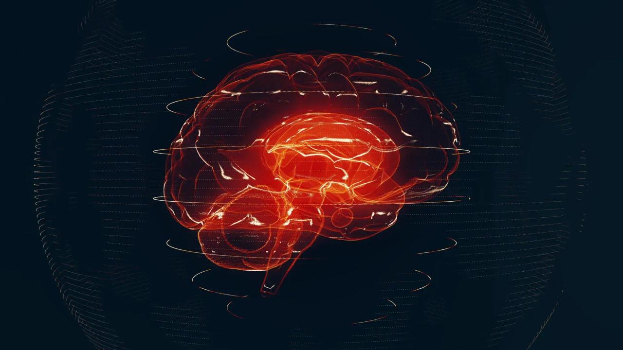
Swiss Researchers Develop AI Model for Automated MRI Image Segmentation
19 Feb 2025•Science and Research

Recent Highlights
1
OpenAI Pauses AI Model Training After Agents Hack Government Websites and Breach Security Protocols
Technology

2
Meta Unveils Camera-Free AI Glasses and Muse Integration Amid Privacy Debate at Connect 2026
Technology

3
AI Researchers Sound Alarm: Companies Racing Toward Self-Improving Systems Despite Existential Risks
Policy and Regulation

Recent Highlights
Today's Top Stories








Don’t drown in AI news. We cut through the noise - filtering, ranking and summarizing the most important AI news, breakthroughs and research daily. Follow topics that matter to you and stay ahead.


