AI-Powered NextBrain Atlas Creates Most Detailed 3D Map of Human Brain for Medical Imaging


4 Sources
[1]
A probabilistic histological atlas of the human brain for MRI segmentation - Nature
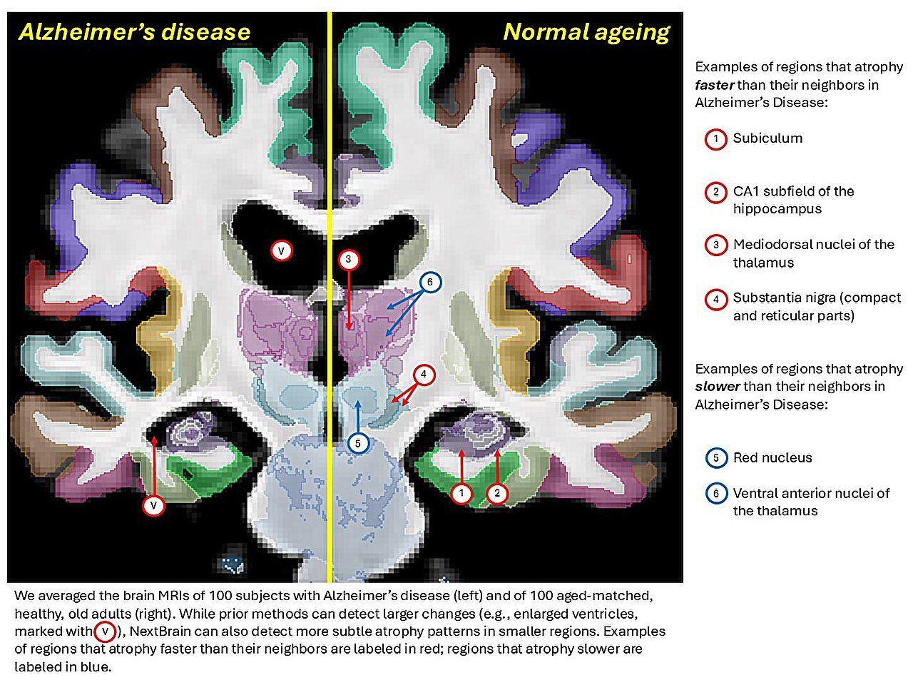
The Bayesian segmentation tool in NextBrain is compatible with 1-mm isotropic scans, as illustrated by the Alzheimer's and ageing experiments. As with other probabilistic atlases, Bayesian segmentation can be augmented with models of pathology to automatically segment pathology, such as tumours55
[2]
New AI-assisted atlas tcan help visualize the human brain in unprecedented detail
University College LondonNov 5 2025 A new AI-assisted brain atlas that can help visualize the human brain in unprecedented detail has been developed by UCL researchers, in a major step forward for neuroscience and neuroimaging. The human brain comprises hundreds of interconnected regions that
[3]
AI Creates the Most Detailed 3D Map of the Human Brain Ever Made - Neuroscience News
Summary: A new AI-powered atlas called NextBrain allows researchers to visualize the human brain in unprecedented detail, down to hundreds of tiny subregions previously invisible on MRI scans. Built from 10,000 microscopic slices of post-mortem brains and aligned with AI, the atlas precisely maps
[4]
New brain atlas offers unprecedented detail in MRI scans
A new AI-assisted brain atlas that can help visualize the human brain in unprecedented detail has been developed by UCL researchers, in a major step forward for neuroscience and neuroimaging. The human brain comprises hundreds of interconnected regions that drive our thoughts, emotions, and
Share
Copy Link
UCL researchers have developed NextBrain, an AI-assisted brain atlas that provides unprecedented detail in MRI scan analysis. The atlas maps 333 brain regions from microscopic tissue data and can analyze living patients' brain scans in minutes, potentially revolutionizing diagnosis of neurological diseases like Alzheimer's.
Revolutionary Brain Mapping Technology
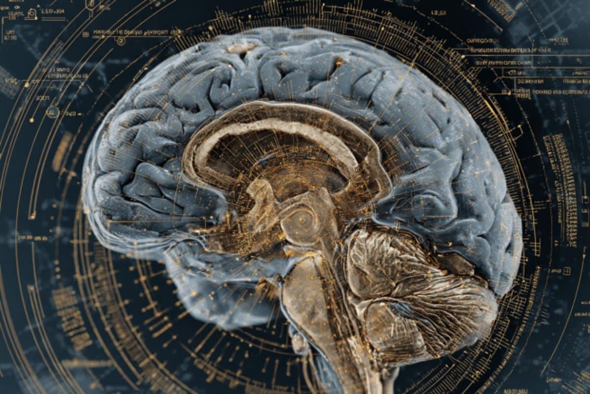
Researchers at University College London (UCL) have developed NextBrain, a groundbreaking AI-assisted brain atlas that represents the most detailed 3D map of the human brain ever created. Published in Nature, this innovative tool enables scientists to analyze MRI scans of living patients with unprecedented precision, identifying hundreds of brain subregions that were previously invisible to conventional imaging techniques
1
2
.
Source: Neuroscience News
The human brain contains hundreds of interconnected regions that control thoughts, emotions, and behaviors. While existing brain atlases can identify major structures like the hippocampus, their finer subregions remain difficult to detect in standard MRI scans. This limitation has significant implications for understanding neurological diseases, as different subregions of brain areas like the hippocampus are affected differently during Alzheimer's disease progression
3
.Six-Year Development Process
The creation of NextBrain required an extraordinary six-year effort that researchers describe as akin to completing a massive jigsaw puzzle. The team used post-mortem tissue from five human brains, each painstakingly dissected and sectioned into approximately 10,000 pieces. These tissue samples were stained to help identify brain structures, photographed under microscopes, and then digitally reassembled into comprehensive 3D models
4
.Before beginning the dissection process, researchers conducted MRI scans of the intact brains to serve as reference images, similar to the picture on a jigsaw puzzle box. AI algorithms played a crucial role in aligning the microscopic images with the MRI scans, accounting for differences between the two imaging techniques while ensuring that reconstructed pieces did not overlap or contain gaps
2
.AI-Accelerated Brain Region Mapping
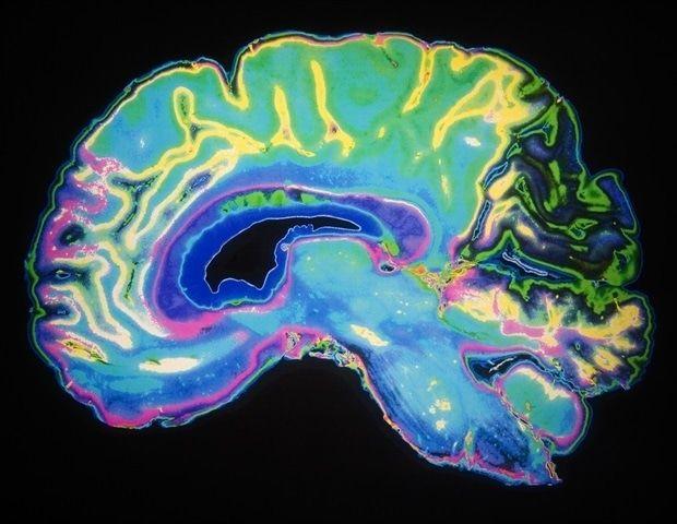
A total of 333 distinct brain regions were labeled on the digital 3D models of each brain, a process that was significantly accelerated by artificial intelligence. According to the researchers, completing this labeling manually would have required decades of work. The resulting atlas represents an average of the five brain models and is generalizable to all adult humans, meaning it can automatically infer detailed information from MRI scans of both living and deceased subjects
3
.
Source: News-Medical
Dr. Juan Eugenio Iglesias, senior author of the study from UCL Medical Physics & Biomedical Engineering and Massachusetts General Hospital/Harvard Medical School, explained that "NextBrain is the culmination of years of effort to bridge the gap between microscope imaging and MRI. By combining high-resolution tissue data with advanced AI techniques, we've created a tool that allows researchers to analyze brain scans at a level of detail that was previously unattainable"
4
.Related Stories
Extensive Validation and Testing
NextBrain underwent rigorous testing on thousands of MRI datasets, demonstrating its ability to reliably identify brain regions across diverse imaging conditions and scanner types. In one validation experiment, the team used the atlas to automatically label brain regions in a publicly available ultra-high-resolution MRI scan, achieving results that closely matched manually labeled regions, even for small areas such as hippocampal subregions
1
.In another significant test, researchers applied NextBrain to over 3,000 MRI scans of living individuals to investigate age-related changes in brain volume. The atlas enabled more detailed analysis of aging patterns than could be achieved using existing neuroimaging tools, demonstrating its superior analytical capabilities
2
.Clinical Applications and Future Impact
The atlas shows particular promise for advancing the diagnosis and treatment of neurological conditions. Dr. Zane Jaunmuktane from UCL Queen Square Institute of Neurology emphasized that NextBrain "provides an unparalleled map of the brain's cellular architecture" that enables "rapid, accurate and accessible analysis of brain images in living individuals, opening the door to detecting the earliest signs of neurological diseases such as Alzheimer's, long before symptoms appear"
4
.
Source: Medical Xpress
The tool's compatibility with various imaging modalities, including 1-mm isotropic scans and ultra-high-field MRI systems, makes it particularly valuable for analyzing high-quality 3D brain images. As advanced imaging technologies become more widely available, NextBrain's ability to extract detailed information from these scans holds significant promise for advancing understanding of the human brain in both health and disease states
1
.All underlying data, tools, and annotations used in NextBrain have been released as open-access resources through the FreeSurfer neuroimaging platform, along with visualization tools and educational materials, ensuring that researchers worldwide can immediately benefit from this breakthrough technology
3
.References
Summarized by

Navi
[3]
[4]
Related Stories
AI-Powered CellTransformer Creates Most Detailed Mouse Brain Map to Date
07 Oct 2025•Science and Research
AI-Powered 3D Brain Mapping Tool Revolutionizes Neurodegenerative Disease Research
20 Mar 2025•Science and Research

New AI Model Maps Regional Brain Aging to Detect Alzheimer's Earlier Than Ever
04 Aug 2026•Science and Research

Recent Highlights
1
Microsoft and Nvidia Launch Surface Laptop Ultra: A $2,599 AI-Ready PC With RTX Spark Chip
Technology

2
Trump Consulted Grok AI Before Ordering Venezuela Invasion and Capture of Nicolás Maduro
Policy and Regulation

3
Scientists Build AI Model That Reconstructs Images From Brain Scans in Record Time
Science and Research
Recent Highlights
Today's Top Stories






Don’t drown in AI news. We cut through the noise - filtering, ranking and summarizing the most important AI news, breakthroughs and research daily. Follow topics that matter to you and stay ahead.


