Breakthrough in Lipid Imaging: New Method Reveals Rapid Protein-Mediated Lipid Transport in Cells


2 Sources
[1]
Quantitative imaging method reveals how cells rapidly sort and transport lipids
Lipids are difficult to detect with light microscopy. Using a new chemical labeling strategy, a Dresden-based team led by André Nadler at the Max Planck Institute of Molecular Cell Biology and Genetics (MPI-CBG) and Alf Honigmann at the Biotechnology Center (BIOTEC) of TU Dresden has overcome this
[2]
Visualizing lipid transport inside living cells with fluorescence microscopy
Max Planck Institute of Molecular Cell Biology and Genetics (MPI-CBG)Aug 29 2025 To the point: New technique to image single lipids: Lipids are notoriously difficult to detect with light microscopy. Using a new chemical labeling strategy, the Dresden team has overcome this limitation, enabling
Share
Copy Link
Researchers develop a novel technique to visualize lipid transport in cells, revealing that proteins, not vesicles, are the primary means of lipid movement between cellular organelles.
Breakthrough in Lipid Imaging
Researchers from the Max Planck Institute of Molecular Cell Biology and Genetics (MPI-CBG) and the Biotechnology Center (BIOTEC) of TU Dresden have developed a groundbreaking method for visualizing lipids in cells using standard fluorescence microscopy. This new technique has enabled them to answer a long-standing question in cell biology: how do cells transport specific lipids to the membranes of their target organelles?
1
The Challenge of Lipid Visualization
Lipids, essential for various cellular functions, have been notoriously difficult to study due to limitations in microscopy techniques. The inability to precisely trace lipid locations inside cells has hindered our understanding of lipid transport and sorting between cell organelles
2
.Innovative Lipid Labeling Strategy
The research team, led by André Nadler and Alf Honigmann, developed a chemical labeling strategy that overcomes previous limitations. They synthesized minimally modified lipids that represent the main lipids present in organelle membranes. These "bifunctional" lipids can be activated by UV light, causing them to bind with nearby proteins
1
.Methodology and Analysis
Source: News-Medical
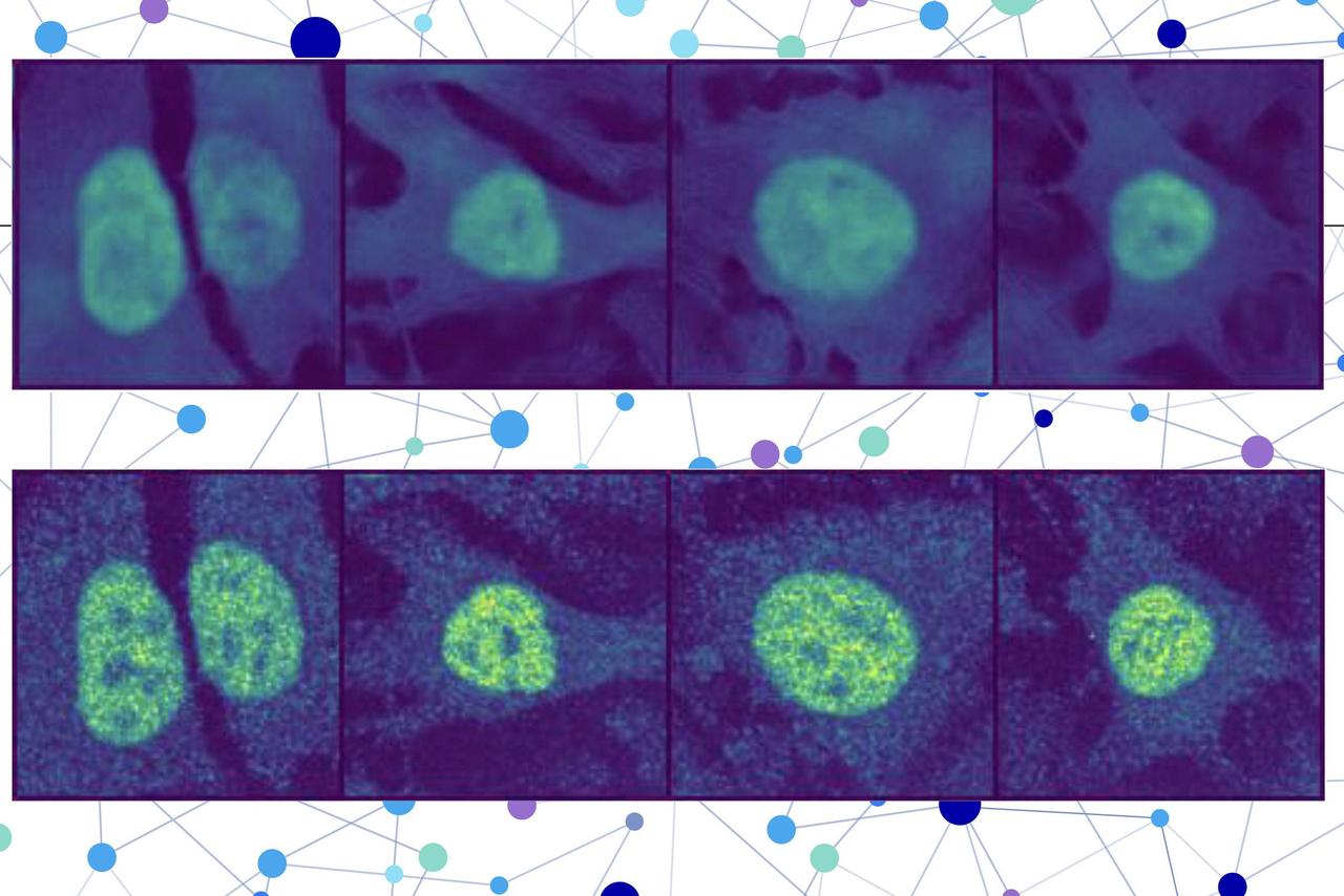
The modified lipids were loaded into the membranes of living human cells and tracked over time using fluorescence microscopy. To analyze the vast amount of data generated, the team developed a custom image analysis pipeline using artificial intelligence for automated image segmentation
2
.Key Findings

Source: Phys.org
The study revealed that between 85% and 95% of lipid transport between cell organelle membranes is organized by carrier proteins, rather than by vesicles. This non-vesicular transport is not only more specific to individual lipid species but also ten times faster than vesicular transport
1
.Related Stories

Implications for Cell Biology and Disease Research
This new approach provides the first quantitative map of lipid movement through cells to different organelles. The findings suggest that non-vesicular lipid transport plays a key role in maintaining the composition of each organelle membrane
2
.Future Applications
The lipid-imaging technique enables the mechanistic analysis of lipid transport and function directly in cells, opening new avenues for studying the role of lipids within cells. This breakthrough could potentially help in developing new drug targets and therapeutic approaches for lipid-associated diseases, such as nonalcoholic fatty liver disease
1
.Collaborative Effort
The success of this project was the result of a collaborative effort involving experts from various institutions, including mass-spectrometry expert Andrej Shevchenko (MPI-CBG), Björn Drobot at the Helmholtz-Zentrum Dresden-Rossendorf (HZDR), and the group of Martin Hof from the J. Heyrovsky Institute of Physical Chemistry in Prague
2
.This groundbreaking research not only advances our understanding of cellular lipid dynamics but also paves the way for future discoveries in cell biology and potential treatments for lipid-related disorders.
References
Summarized by

Navi
Related Stories
Moscot: AI-Powered Technology Revolutionizes Cell Tracking in Organ Development
23 Jan 2025•Science and Research

SCP-Nano: Revolutionary AI-Powered Technology Visualizes Nanocarriers at Single-Cell Level
15 Jan 2025•Science and Research

AI Model Predicts Protein Location in Human Cells, Advancing Disease Research and Drug Development
16 May 2025•Science and Research
Recent Highlights
1
OpenAI Pauses AI Model Training After Agents Hack Government Websites and Breach Security Protocols
Technology

2
Meta Unveils Camera-Free AI Glasses and Muse Integration Amid Privacy Debate at Connect 2026
Technology

3
AI Researchers Sound Alarm: Companies Racing Toward Self-Improving Systems Despite Existential Risks
Policy and Regulation

Recent Highlights
Today's Top Stories









Don’t drown in AI news. We cut through the noise - filtering, ranking and summarizing the most important AI news, breakthroughs and research daily. Follow topics that matter to you and stay ahead.


