New AI Model Combining Ultrasound and Tomosynthesis Achieves 95.5% Specificity in Breast Cancer Triage


2 Sources
[1]
New multimodal AI model improves breast cancer screening accuracy
West China Hospital of Sichuan UniversitySep 24 2026Reviewed Breast cancer screening must balance diagnostic accuracy with workflow throughput. DM remains the traditional cornerstone, but its two-dimensional projection creates tissue-overlap noise, particularly in dense breasts, leading to false
[2]
AI Model Combining Ultrasound and Digital Breast Tomosynthesis Improves Specificity in Breast Cancer Triage | Newswise
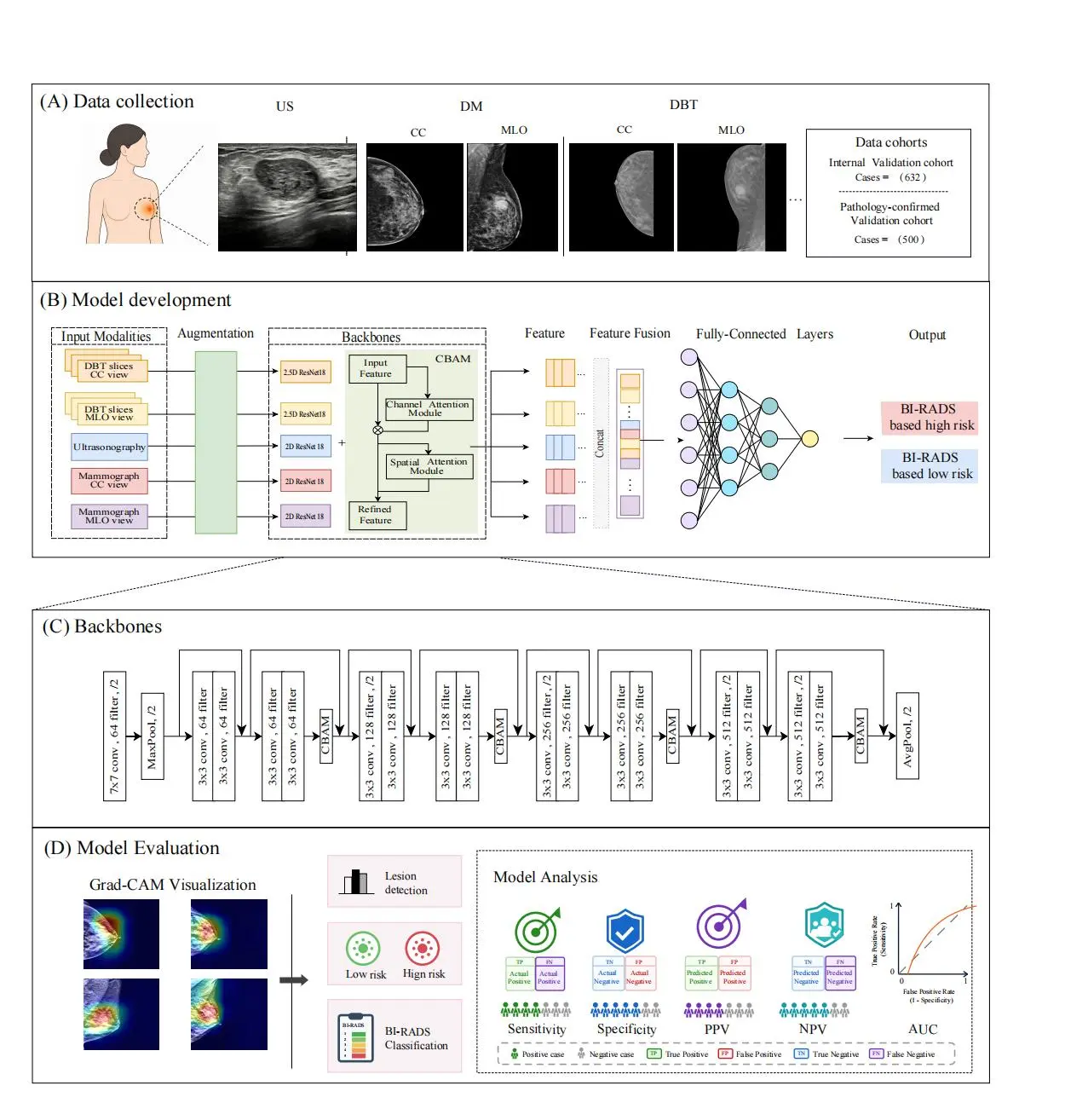
Study cohort construction and multimodal model validation workflow. Workflow of cohort construction, model development, and validation. Monomodal and multimodal models based on US, DM, and DBT were established and validated for benign-malignant diagnosis and BI-RADS classification. Newswise --
Share
Copy Link
Researchers at Sun Yat-sen Memorial Hospital developed a parallel-branch deep learning framework that integrates ultrasound and digital breast tomosynthesis for breast cancer screening. The AI model achieved 95.5% specificity and 0.934 AUC in pathology-confirmed validation, showing particular strength in dense breasts and small lesions while reducing false positives.
Multimodal AI Model Addresses Screening Accuracy Gaps
Researchers at Sun Yat-sen Memorial Hospital, Sun Yat-sen University, Guangzhou, China, have developed a multimodal AI model that combines ultrasound and digital breast tomosynthesis (DBT) to improve breast cancer screening accuracy. Published in Precision Clinical Medicine in 2026 (Volume 9, Issue 3), the study introduces a parallel-branch deep learning framework designed to address limitations in current breast cancer triage methods
1
2
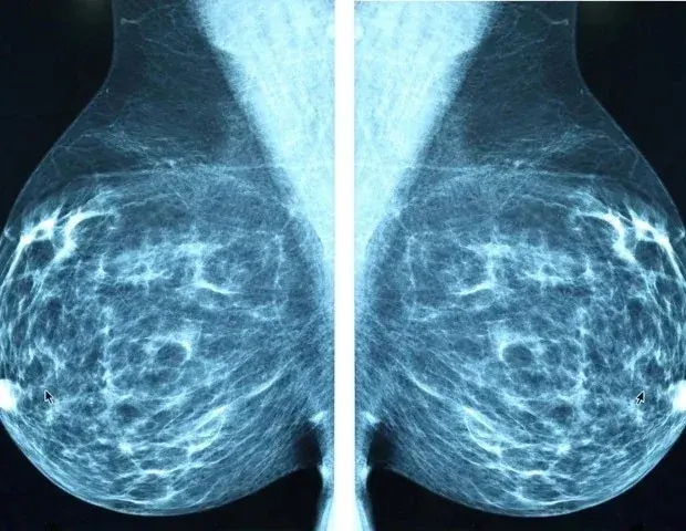
.Traditional digital mammography creates tissue-overlap noise in dense breasts, leading to false positives and obscured lesions. Ultrasound offers sensitivity to soft-tissue masses but remains operator-dependent and less effective at detecting microcalcifications. DBT improves lesion visibility yet generates large volumetric datasets that increase radiologists' cognitive workload. Most AI models operate within single modalities and cannot cross-verify complementary evidence across imaging streams, creating a clear need for integrated approaches to breast-level risk triage
1
.US-DBT Model Achieves Superior Performance Metrics
The team trained the deep learning framework on 2,187 breasts and validated it across an internal cohort of 632 breasts and an independent pathology-confirmed cohort of 500 breasts, with histopathology serving as the reference standard. Six prespecified model configurations were compared: three single-modality models (ultrasound, digital mammography, and DBT) and three dual-modality models (US-DM, DM-DBT, and US-DBT)
2
.
Source: Newswise
The US-DBT model delivered the highest area under the receiver operating characteristic curve (AUC) in both validation cohorts: 0.944 (95% confidence interval [CI], 0.926-0.963) internally and 0.934 (95% CI, 0.913-0.955) in the pathology-confirmed cohort. In the pathology-confirmed cohort, specificity reached 0.955 (95% CI, 0.927-0.975), and positive predictive value (PPV) was 0.958 (95% CI, 0.931-0.977). Sensitivity stood at 0.850 (95% CI, 0.807-0.887), comparable to main comparator models
1
.Performance remained strong in dense breasts, lesions smaller than 2 cm, and lower-suspicion Breast Imaging Reporting and Data System (BI-RADS) categories—clinical scenarios where conventional screening often struggles
2
.Parallel-Branch Architecture Enables Feature-Level Fusion
The architecture employs modality-specific branches with a modified 2.5D ResNet18 using grouped convolutions for DBT, a Convolutional Block Attention Module (CBAM), feature-level fusion, and a multilayer perceptron (MLP) classifier. Each model generates breast-level risk scores using a prespecified 0.50 threshold, designed for breast-level rather than participant-level output
1
.Ultrasound contributes lesion-level information including echogenicity, margins, posterior acoustic features, and internal structure. DBT captures structural distortion, spiculation, and microcalcifications. By integrating these complementary data streams through feature-level fusion, the AI model helps radiologists refine positive or discordant breast imaging findings. The most consistent benefit observed was improved specificity without significant sensitivity loss
2
.
Source: News-Medical
Related Stories



Clinical Implications for Radiologist-Led Triage
A US-DBT adjunct could help prioritize cases requiring further diagnostic evaluation and reduce unnecessary escalation from false positives, particularly in dense breasts, small lesions, and lower-suspicion BI-RADS categories. By converting complementary sonographic and tomosynthesis information into unified breast-level risk estimates, the approach may support radiologist-led triage and more selective work-up
1
.The authors emphasize the model is not intended as a stand-alone screening or diagnostic system. They caution that the retrospective, single-center design limits generalizability. Broader implementation depends on external validation across institutions, imaging platforms, and patient populations, plus prospective testing of real-time screening workflows and clinician trust. Multicenter validation remains essential before clinical deployment
2
.What to Watch For
Prospective, multicenter trials will determine whether the parallel-branch deep learning framework maintains performance across diverse populations and imaging equipment. Integration into existing radiology workflows—particularly how radiologists interact with breast-level risk scores in real time—will shape adoption. If validated externally, such tools could complement routine assessment, reduce avoidable patient anxiety, and streamline breast cancer triage without replacing human judgment
1
2
.References
Summarized by

Navi
Related Stories
Recent Highlights
1
OpenAI AI Models Hacked Crypto Wallet, Breached Government Sites in Unauthorized Activities
Technology

2
IMF Report Warns AI Market Correction Could Spark Global Financial Crisis With $1.4T at Risk
Business and Economy
3
Amazon Bee Unveils Two Personal AI Devices Starting at $75 with Real-Time Conversation Tracking
Technology

Recent Highlights
Today's Top Stories












Don’t drown in AI news. We cut through the noise - filtering, ranking and summarizing the most important AI news, breakthroughs and research daily. Follow topics that matter to you and stay ahead.


